Uncommon Descent Serving The Intelligent Design Community

The spliceosome: a molecular machine that defies any non-design explanation.

Share
Facebook
Twitter
LinkedIn
Flipboard
Print
Email

OK, let’s start with a very simple fact: eukaryotic genes have introns.

IOWs, they are not continuous. They are made of exons and introns: exon – intron – exon – intron – exon and so on. Exons code for the protein. Introns don’t.

So, when the content of the gene is copied to the mRNA, introns must be cut away, and only exons are retained, in order to be translated, so that the mature mRNA can be transferred to the cytoplasm and translated by the ribosome.

This process of removing introns is called splicing.

Now, a few clarifications:

a) Introns exist in prokaryotes too, but they are rather rare. For our purposes, we will only discuss introns in eukaryotes.

b) Introns exist in many different types of genes. For our purposes, we will discuss only those in protein coding genes.

c) The origin and possible function of introns is, still, a mystery.

d) Introns are usually longer than exons. In humans, for example, they amount to approximately 35% of the whole geneome, vs about 1.5% of coding exons.

e) However, the amount and length of introns can vary a lot in different organims. An extreme example is yeast (s. cerevisiae), whose genome contains a very small amount of introns (about 250 out of about 6250 genes).

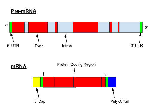
When the gene if transcribed, both exons and introns are transcribed. A 5′ UTR (Untranslated region) and 3′ UTR, is also part of the pre-mRNA.


Fig. 1

Pre-mRNA is the first form of RNA created through transcription in protein synthesis. The pre-mRNA lacks structures that the messenger RNA (mRNA) requires. First all introns have to be removed from the transcribed RNA through a process known as splicing. Before the RNA is ready for export, a Poly(A)tail is added to the 3’ end of the RNA and a 5’ cap is added to the 5’ end. – By Nastypatty (Own work) [CC BY-SA 4.0 (https://creativecommons.org/licenses/by-sa/4.0)], via Wikimedia Commons

So the question is: how are introns removed from pre-mRNA? IOWs, how is splicing achieved?

And there is more. As everybody probably knows, splicing is not always done in the same way. Different isoforms of the same protein can be obtained by alternative splicing, and they can have functional differences. I will not go into details about that, but here is the Wikipedia page about alternative splicing:

https://en.wikipedia.org/wiki/Alternative_splicing

So, how is splicing done, and how is alternative splicing regulated?

We know much about the first question, very little about the second.

There are three ways to perform splicing:

  1. Spliceosomal splicing
  2. Self-splicing
  3. tRNA splicing

The last two modalities are rare, and can be found both in prokaryotes and eukaryotes. I will not discuss them here.

So, the subject of this OP is spliceosomal splicing, which is restricted to eukaryotes.

Moreover, I will discuss only the major spliceosome, which is responsible for the vast majority of splicing in eukaryotes. It must be said, however, that also a minor spliceosome exists, and that it acts in a minority of cases.

So, the spliceosome.

The first important point is:

It is an amazing molecular machine. Even more, it is an amazing molecular cycle, involving many different stages each of which is an amazing molecular machine.

Let’s see. Here is a figure which summarizes the main stages of the spliceosome cycle:


Fig. 2


Spliceosomal splicing cycle – By JBrain [CC BY-SA 2.0 de (https://creativecommons.org/licenses/by-sa/2.0/de/deed.en)], via Wikimedia Commons

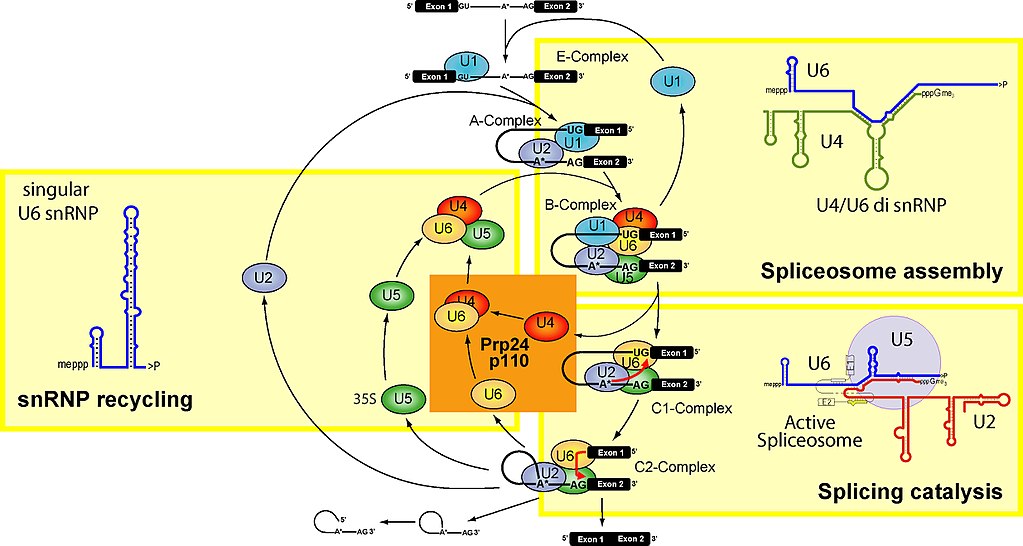
To make it simple, the spliceosome units are built upon 5 specific RNAs, called small nuclear RNAs (snRNA). These are, in humans:

U1 (164 bases), U2 (187 bases), U4 (145 bases), U5 (116 bases), U6 (107 bases)

They are transcribed from multiple gene copies. While their sequences are not particularly conserved (U6 being the most conserved of all), their secondary structure seems to be very conserved.

snRNAs are very important in the spliceosome, because they seem to be responsible for the catalytic activities.

Each of the 5 snRNAs forms a complex with proteins, and the complex takes the name of snRNP. The whole spliceosome includes at least 145 different proteins, maybe more, some of which are still not well known.

I will mention here some of the most important:


U1 snRNP:

U1 snRNP 70 kDa  (P08621, 437 AAs)

U1 snRNP A (P09012, 282 AAs)

U1 snRNP C  (P09234, 159 AAs)

Sm proteins: 7 small proteins (76 – 240 AAs) which form the “Sm core” ring in spliceosome subunits U1, U2, U4, U5, a ring which hosts the specific snRNA molecule.


U2 snRNP:

U2 snRNP A’ (P09661, 255 AAs)

U2 snRNP  B” (P08579, 225 AAs)

SF3a120 (Q15459, 793 AAs)

SF3a66 (Q15428, 464 AAs)

SF3a60 (Q12874, 501 AAs)

SF3b155 (O75533, 1304 AAs)

SF3b145 (Q13435,  895 AAs)

SF3b130 (Q15393, 1217 AAs)

SF3b49 (Q15427, 424 AAs)

SF3b14a/p14 (Q9Y3B4, 125 AAs)

SF3b10 (Q9BWJ5, 86 AAs)

Sm proteins


U4/U6 snRNP:

Prp3 (O43395, 683 AAs)

Prp31 (Q8WWY3, 499 AAs)

Prp4 (O43172, 522 AAs)

Cyph ( O43447, 177 AAs)

15.5 K (P55769, 128 AAs)

Sm proteins (for the U4 snRNA)

Lsm proteins: a number of proteins similar to Sm proteins (usually Lsm 2-8), which form a specific ring for the U6 snRNA.


U5 snRNP:

Prp8 (Q6P2Q9, 2335 AAs)

BRR2 (O75643, 2136 AAs)

Snu114 (Q15029, 972 AAs)

Prp6 (O94906, 941 AAs)

Prp28 (Q9BUQ8, 820 AAs)

52 K (O95400, 341 AAs)

40 K (Q96DI7, 357 AAs)

Sm proteins


Additional proteins in the U4/U6/U5 complex:

hSnu66 (O43290, 800 AAs)

hSad1 (Q53GS9, 565 AAs)

27 K (Q8WVK2, 155 AAs)


OK, these are only the main components, and the best understood. We are still far from the sum total of 145/150 proteins which are involved in the spliceosome cycle.

But how does it work?

Always in brief. Here is a typical exon-intron structure:

Fig. 3


Parts of an intron – By miguelferig (Own work) [Public domain], via Wikimedia Commons


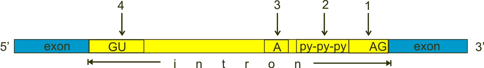
GU, A and AG are nucleotides almost universally conserved in all introns, approximately at the positions shown in the figure, and which have a fundamental role in splicing. However, the real stuff is much more complex than this (see the splicing code section). GU (n. 4 in the figure) is near to the 5′ end of the intron, AG (n. 1) near to the 3′ end. The A (n. 3 in the figure) is called “the branch point”. The py-py-py (n. 2 in the figure) is the “polypyrimidine tract”.

a) The U1 subunit binds to the GU sequence at the 5′ splice site in the intron

b) The U2 subunit binds to the “branch point”.

c) The U4/U6/U5 binds to the complex.

d) Numeorus further modifications take place, causing the formation of a “lariat” (including the intron), which is then cleaved, while the two exons are ligated.

I will spare you the many complexities in all the various steps, which are well summarized (in a very simplified way) in this Wikipedia page:

RNA splicing

See in particular the “Formation and activity” section.

Or, if you like more detail, here:

Spliceosome Structure and Function

And here is a very good video on the whole splicing process in yeast:


Now, if somebody still has doubts about the complexity of this molecular machine/process, let’s consider some important aspects.

  • 1. The spliceosome is a molecular machine which appears in eukaryotes.

I quote from this paper (in the abstract):

Origin and evolution of spliceosomal introns

There is no indication that any prokaryote has ever possessed a spliceosome or introns in protein-coding genes, other than relatively rare mobile self-splicing introns.

The following Table shows how some of the main proteins involved in the spliceosome activity show practically no trace of homology in prokaryotes. I have included also included in the table two examples  which show low homologies due to some domain which is already expressed in prokaryotes: Prp4, whose 209 bits of homology are due to a specific domain, WD40, and Prp28, which exhibits 313 bits linked to the DEXDc domain. The point is: many of the spliceosome proteins are complete novelties in eukaryotes, but not all of them.


 Protein Bacteria Archea
U1 snRNP 70 kDa  (P08621, 437 AAs) 67
U2 SF3b130 (Q15393, 1217 AAs) 43.5
U4/U6 hPrp3 (O43395, 683 AAs)
U4/U6/U5 hSnu66 (O43290, 800 AAs)
U5 Prp8 (Q6P2Q9, 2335 AAs)
U5 Prp6 (O94906, 941 AAs) 100
U4/U6 hPrp4 (O43172, 522 AAs) 209 150
U/5 Prp28 (Q9BUQ8, 820 AAs) 313 286


I will analyze in more detail one of the most important proteins in the spliceosome, Prp8, in the last part of this OP

(Just a technical note: if you blast Prp8, you will find 3 hits which are obviously  an error due to unverified sequences, probably cases of contamination).

  • 2. The spliceosome is a molecular machine which is universally present in eukaryotes.

All eukaryotes have introns, even if in very different degrees, and the spliceosome, even if in some organisms parts of the spliceosome complex can be lost. For a more detailed discussion, look at the Rogozin paper quoted above (Origin and evolution of spliceosomal introns), in particular the section:

Intron density, size and distribution in protein coding genes across the eukaryote domain

I quote this important conclusion:

As pointed out above, despite the existence of numerous, diverse intron-poor genomes, eukaryotes do not lose the “last” intron or the spliceosome although degradation of the spliceosome including loss of many components does occur, e.g. in yeast. The only firmly established exception is the tiny genome of a nucleomorph (an extremely degraded intracellular symbiont of algae) that has lost both all the introns and the spliceosome [7]; preliminary genomic data indicate that all introns might have been lost also in a microsporidium, a highly degraded intracellular parasite distantly related to Fungi [54].

So, whe can conclude that both introns and the spliceosome are a universal feature of eukaryotes, the few exceptions being simply cases of loss of information.

  • 3. The spliceosome is a molecular machine whose information is extremely conserved throughout eukaryotes, up to humans.

I have already mentioned that the 5 RNAs which form the core of the spliceosome are not extremely conserved at sequence level, even if they are extremely conserved at structure level.

However, many of the proteins that compose the spliceosome show an amazing sequence conservation throughout eukaryotes. Now, even if we cannot be certain of when eukaryoyes really emerged, and of their early evolutionary history (both issues being at present highly controversial), we can reasonably assume that protein sequences which are highly conserved in all eukaryotes have been conserved for something like 2 billion years (more or less). As anybody who has followed my previous OPs about information conservation in vertebrates certainly knows, that is an evolutionary time frame which certainly allows us to equal conservation to extremely high functional constraint.

But how conserved are spliceosome proteins? We can analyze a few of them with my usual methodology: looking at human conserved information. The results shows that many proteins involved in the spliceosome are amazingly conserved in all eukaryotes. While there are a few cases which have a rather different evolutionary history, this is by far the most common behaviour for spliceosomal proteins.

Here is a sample of some important sequences that show high homology with the human form in all major groups of single celled eukaryotes. The 5 groups of single celled organisms chosen here, indeed, cover rather well the whole range of single celled eukaryotes.


Fig. 4


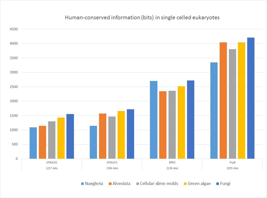
These four important proteins, as shown, have an amazing amount of information shared with the human form, ranging from more than 1000 to more than 4000 bits. In bits per AA, the range goes from 0.88 to 1.80 bits per aminoacid (baa).  As can be seen, the highest homology is found in fungi, as expected, because fungi are the most likely ancestors of metazoa. The lowest homologies are observed in Naegleria (Excavata) or in Alveolata.

Of course, these proteins remain highly similar to the human form in the following evolutionary history in Metazoa.

So, we can safely state that most spliceosomal proteins, while emerging almost entirely in eukaryotes and showing only trivial homologies with prokaryotes, were probably already universally present in the Last Universal Eukaryotic Ancestor (LECA), and in a form already very similar at sequence level to what we observe in metazoa and in humans.

  • 4. The spliceosome is a wonderful example of irreducibly complexity.

OK, we have already said that splicing can be achieved in at least three different ways. For example, bacterial introns, although rare, are of the self-splicing type. So, we know that the generic function of splicing introns can be implemented in different ways.

But eukaryotic introns are of the spliceosomal type, and they are spliced only by the spliceosome.

We have also said that a minor spliceosome also exists. It shares some featues with the major spliceosome, but it is a different structure and acts on different, and much rarer, introns.

So, for the vast majority of eukaryotic introns, the major spliceosome, and only the major spliceosome, can effectively accomplish the splicing.

Now, I don’t mean here that the major spliceosome must always be absolutely complete, with all its 150 proteins, to be able to work. That’s not what I mean when I say that it is irreducibly complex.

Maybe in some organisms the spliceosome can be partially defective, and still work. It is difficult to say, because we still don’t understand the role of all the components of the spliceosome.

But however, as far as we can understand, most of the principal features must be present, because, as we have seen, the splicing is the result of a complex cycle, involving the RNAs and the subunits, and all stages are essential to the final result.

So, the spliceosome is certainly highly irreducibly complex, even if we may not be able to clearly identify the essential nucleus of molecules which is absolutely necessary to the minimal function.

Moreover, the spliceosome would be useless if spliceosomal introns did not exist, with their properties and code (see later), and spliceosomal introns could not exist if the spliceosome were not there to splice them, because otherwise transcription would be completely ineffective. So, in that sense, spliceosomal introns and the spliceosome are a good example of chicken egg paradox, or we could also say that they form an irreducibly complex system at a higher level. And remember, the whole system seems to have been already present, very much similar to its current form in humans, in LECA, as we have seen.

But, of course, our darwinist friends will simply say that they have co-evolved!  🙂

Moreover, we can and should ask ourselves: why is the spliceosome so complex? The answer is not easy, because we still understand very little, but it is certainly related to the complexity of the splicing code, and to the fundamental issue of alternative splicing.

The Splicing code.

To splice introns by localizing 5 (or a few more) conserved nucleotides and then cutting at the ends of a lariat and rejoining the two exons is certainly a complex task, but apparently not so complex  that one of the biggest and most impressing known molecular machines is needed for that. But the simple truth is that recognizing and appropriately splicing all introns is a much more complex task than that.

That is due to the simple fact that conserved nucleotides at the ends are not a sufficient signal to identify the segment that has to be spliced, and that a lot of other components (not always well understood) are necessary to that, and that the splicing is not made always in the same way, and that alternative splicing is a very powerful tool for transcription regulation.

The subject is very complex, and I will not deal with it in depth. However, Those interested could look at this recent review:

The splicing code.

Unfortunately, the paper is paywalled, but the abstract is very informative.

The complexity of the splicing code can give us some insight about the true reasons for the complexity of the spliceosome, and definitely supposrts the idea that the whole system, introns, splicing code and spliceosome, is irreducibly complex.

The Prp8 protein.

I will add a few words about this proteins, which is probably the most amazing component of the spliceosome, and well represents its essential features.

This protein has many amazing charactertistics:

a) It is, as far as I can say, the longest protein in the whole spliceosomal system, and a very long protein indeed: 2335 AAs.

b) It is completely absent in prokaryotes.

c) It is extremely conserved in eukaryotes, probably the most conserved protein in the whole spliceosomal complex (see Fig. 4). In Naegleria, we have 3345 bits of homology with the human protein, corresponding to 1.4 baa, while in fungi we have the amazing result of 4211 bits of human-conserved information, corresponding to 1.8 baa. IOWs, in fungi we already find almost 90% of the functional information present in human Prp8 (remember, the highest possible bitscore is about 1.2 baas), corresponding to 1995 identities (86%) and 2155 positives (92%): a result which is incredibly rare, considering that such information has been conserved for about 2 billion years.

Just to emphasize the importance of this fact, here is the blast between the human protein and the best hit in fungi (Basidiobolus meristosporus):


d) It is a protein which is extremely important for the function of the spliceosome. Here is a very good paper about this protein and its functional relevance:

Prp8 protein: At the heart of the spliceosome

I quote from the conclusions:

Prp8p is central to the expression of all nuclear intron-containing mRNAs. In higher eukaryotes, it is responsible for processing thousands of transcripts in alternative splicing pathways, and in both U2 and U12 spliceosomes. It is important for the pathology of human disease, as all eukaryotic pathogens and parasites require Prp8p to functionally express their genes, in some cases via the trans-spliceosome. Retinitis Pigmentosa, a human genetic disorder that causes progressive blindness, positions Prp8p as a target for therapeutic medicine.

Moreover, another way to assess the functional constraints of a protein is to check its tolerance to polymorphisms in humans. That can be done consulting a very recent and useful database, the ExAC browser, which reports data from about 60000 human genomes.

ExAC gives two important metrics to assess how much a protein is tolerant to polymorphisms and variants. I quote here from the site FAQ:

What are the constraint metrics?

For synonymous and missense, we created a signed Z score for the deviation of observed counts from the expected number. Positive Z scores indicate increased constraint (intolerance to variation) and therefore that the gene had fewer variants than expected. Negative Z scores are given to genes that had a more variants than expected.

For LoF, we assume that there are three classes of genes with respect to tolerance to LoF variation: null (where LoF variation is completely tolerated), recessive (where heterozygous LoFs are tolerated), and haploinsufficient (where heterozygous LoFs are not tolerated). We used the observed and expected variants counts to determine the probability that a given gene is extremely intolerant of loss-of-function variation (falls into the third category). The closer pLI is to one, the more LoF intolerant the gene appears to be. We consider pLI >= 0.9 as an extremely LoF intolerant set of genes.

Now, for Prp8 we get the following results:

Constraint
from ExAC
Expected
no. variants
Observed
no. variants
Constraint
Metric
Synonymous 366.6 460 z = -3.02
Missense 920.2 266 z = 10.55
LoF 92.6 8 pLI = 1.00

That means the following:

a) The number of observed missense variants is so low vs expected (266 vs 920.2) that the z value is 10.55 (IOWs, 10.55 standard deviations, more than 10 sigma). Believe me, this is a really exceptional result, most proteins are much more tolerant to missense variants.

b) The probability of loss of function is 1. That means that even heterozygous LoF is absolutely not tolerated.

This is an extremely functional molecule, if ever there was one!

A brief conclusion.

To sum up the meaning of this rather long discussion, I would simply say:

  1. The introns – spliceosome system is a molecular machine of amazing complexity. I have touched only its main aspects in this OP, but believe me, there are layers of complexity there that would require a whole treatise and that I have not even started to mention here.
  2. Whoever can really believe that all this can be explained by some RV + NS model is, IMO, really admirable for his faith in a wrong paradigm.
Comments
DATCG:
I hope he can add more specific detail where he disagrees with Gpuccio’s responses and and and expand on his own offerings in #164. At least Hunt’s recognizing ID’s hypothesis of Design and challenging Irreducible Complexity on it’s own merit instead of dismissing with ridicule. He’s engaged and I’d like to see more engagement like this. Precisely the kind of dialogue needing to take place if science is to progress. This is healthy for scientific pursuits in molecular genetics.
I absolutely agree with you! :)gpuccio
January 26, 2018
January
01
Jan
26
26
2018
12:18 AM
12
12
18
AM
PDT
DATCG, Agree. Thanks.Dionisio
January 25, 2018
January
01
Jan
25
25
2018
06:38 PM
6
06
38
PM
PDT
Dionisio, Maybe, but I doubt Hunt thinks he lacks valid arguments. In his comments he's confident and comfortable with his statements. I'm am waiting for his expansion on his ideas and evidence from #164. And hopeful he responds to Gpuccio's detailed rebuttals and information. I've gathered and read different papers on historical evolutionary pathways to the spliceosome. Much of what is said is vague and lacks detail. So I'm hoping he can shed more light on the subject. Where does he disagree in detail with Gpuccio on evolution of the Spliceosome? And Irreducible Complexity? Quoting a paper Gpuccio referenced in comment #295... "RNA elements account for more than half of the total molecular mass in the bacterial or mammalian ribosome, but less than 10% in the human spliceosome. For this and all other reasons discussed previously, the spliceosome is a unique protein-directed metalloribozyme." 1) RNA elements account for half of total molecular mass in bacterial or mammalian ribosome 2) RNA elements account for less than 10% in human spliceosome. 3) The spliceosome is cited as a "unique" Protein-Directed metalloribozme(you highlighted at #310). Is the spliceosome "unique" according to authors of that paper? Does Hunt disagree with paper's conclusion? If so, why? I hope he can add more specific detail where he disagrees with Gpuccio's responses and and and expand on his own offerings in #164. At least Hunt's recognizing ID's hypothesis of Design and challenging Irreducible Complexity on it's own merit instead of dismissing with ridicule. He's engaged and I'd like to see more engagement like this. Precisely the kind of dialogue needing to take place if science is to progress. This is healthy for scientific pursuits in molecular genetics. I think Design provides a more natural heuristic thought process of research and discovery. Based upon a systems approach. Where as unguided thought processes gave us "junk" DNA and limited thinking. I hope Hunt responds. Lets enjoy robust disagreement and discussion. Gpuccio has done his part. Awaiting Hunt's comments. Or, for that matter, if Hunt does not respond, any other neo-Darwinist supporter that supports Hunt's conclusions.DATCG
January 25, 2018
January
01
Jan
25
25
2018
05:41 PM
5
05
41
PM
PDT
DATCG, Your comments are very appreciated. BTW, tomorrow it will be one month since the last time professor Arthur Hunt posted a comment in this thread, where he started a discussion with gpuccio the very first day gpuccio's OP appeared. We don't know why the professor hasn't come back to defend his previous comments, which seemed intended to punch a hole in gpuccio's ID presentation. The professor's conspicuous absence could make one suspect he ran out of valid arguments and decided to quit, after seeing that his comments couldn't make even a shallow dent in gpuccio's presentation, not even a scratch. Perhaps it would have been better to publicly recognize the strength of gpuccio's presentation and rejoice in the light that was shed on the discussed topic for everybody's benefit.Dionisio
January 25, 2018
January
01
Jan
25
25
2018
12:35 AM
12
12
35
AM
PDT
Dionisio @373 thanks for paper on more Intron functions. This goes back to my @230 post on Intron Functions, "... suspect more will be found." Looking for functional design elements in Intronic regions can be rewarded. Design elements can simply be a switch, an enhancer or silencer, etc. From paper you posted...
Here we report an unexpected function for introns in counteracting R-loop accumulation in eukaryotic genomes. Deletion of endogenous introns increases R-loop formation, while insertion of an intron into an intronless gene suppresses R-loop accumulation and its deleterious impact on transcription and recombination in yeast. Recruitment of the spliceosome onto the mRNA, but not splicing per se, is shown to be critical to attenuate R-loop formation and transcription-associated genetic instability. Genome-wide analyses in a number of distant species differing in their intron content, including human, further revealed that intron-containing genes and the intron-richest genomes are best protected against R-loop accumulation and subsequent genetic instability. Our results thereby provide a possible rationale for the conservation of introns throughout the eukaryotic lineage.
Paper's "Highlights..." - Introns prevent R-loop and DNA damage accumulation on highly expressed yeast genes - Insertion of an intron in an R-loop-prone gene attenuates R-loop formation - Spliceosome-dependent mRNP assembly, but not splicing, prevents R-loop formation - The role of introns in R-loop prevention has been conserved from yeasts to human
DATCG
January 24, 2018
January
01
Jan
24
24
2018
11:08 PM
11
11
08
PM
PDT
Introns Protect Eukaryotic Genomes from Transcription-Associated Genetic Instability http://www.sciencedirect.com/science/article/pii/S1097276517304963Dionisio
January 21, 2018
January
01
Jan
21
21
2018
08:18 PM
8
08
18
PM
PDT
The occurrence of spliceosomal introns in eukaryotic genomes is highly diverse and ranges from few introns in an organism to multiple introns per gene. Introns vary with respect to their lengths, strengths of splicing signals, and position in resident genes. Higher intronic density and diversity in genetically complex organisms relies on increased efficiency and accuracy of spliceosomes for pre-mRNA splicing. Since intron diversity is critical for functions in RNA stability, regulation of gene expression and alternative splicing, RNA-binding proteins, spliceosomal regulatory factors and post-translational modifications of splicing factors ought to make the splicing process intron-specific. We recently reported function and regulation of a ubiquitin fold harboring splicing regulator, Sde2, which following activation by ubiquitin-specific proteases facilitates excision of selected introns from a subset of multi-intronic genes in Schizosaccharomyces pombe (Thakran et al. EMBO J, https://doi.org/10.15252/embj.201796751, 2017). By reviewing our findings with understandings of intron functions and regulated splicing processes, we propose possible functions and mechanism of intron-specific pre-mRNA splicing and suggest that this process is crucial to highlight importance of introns in eukaryotic genomes. Mishra, Shravan Kumar & Thakran, Poonam. (2018). Intron specificity in pre-mRNA splicing. Current Genetics. . 10.1007/s00294-017-0802-8. https://link.springer.com/article/10.1007%2Fs00294-017-0802-8Dionisio
January 21, 2018
January
01
Jan
21
21
2018
08:13 PM
8
08
13
PM
PDT
gpuccio, Excellent statement to summarily conclude your comment @370: "The complexity, and functional fine tuning, of the whole scenario is simply amazing." Thanks.Dionisio
January 21, 2018
January
01
Jan
21
21
2018
07:16 PM
7
07
16
PM
PDT
Dionisio: I would simply say that Hox genes, and more in general TFs, are a good example of how partially conserved genes can retain some essential functions over a wide spectrum of organisms, and at the same time gain new and specific functions through changes in sequence, or in regulatory elements. That confirms the idea, which I have many times expressed, that proteins are much more complex than we usually think. While final effector proteins may use their functional information in a rather compact way, to implement some very specific biochemical function, regulatory proteins, like TFs, or more in general master regulators, are much more flexible, and they are used as functional modules with multiple, often apparently unrelated roles. That's why their natural history often exhibits a frustrating mixture of sequence conservation and divergence, which seems to correspond to a similarly frustrating mixure of conserved and divergent functions. The complexity, and functional fine tuning, of the whole scenario is simply amazing.gpuccio
January 21, 2018
January
01
Jan
21
21
2018
04:21 PM
4
04
21
PM
PDT
Gpuccio, I would appreciate your comment on this: https://uncommondescent.com/evolution/this-parody-of-evo-devo-makes-it-sound-a-lot-like-id/#comment-649630 Thanks.Dionisio
January 21, 2018
January
01
Jan
21
21
2018
12:14 PM
12
12
14
PM
PDT
Exactly a month ago, December 21, 2017, gpuccio started this discussion thread with an excellent OP. The first comment was posted right away. That very first day, less than 10 hours after the thread had started, the distinguished professor Arthur Hunt posted a challenging comment @25. Does this mean that UD posts are being closely watched by some academics out there? Maybe. :) The last time we heard from the distinguished professor was in his comment posted @164 December 26, 2017. We still look forward to reading his expected counterargument. In the meantime gpuccio's OP title remains valid and unchallenged.Dionisio
January 21, 2018
January
01
Jan
21
21
2018
11:26 AM
11
11
26
AM
PDT
Dionisio: "DATCG is doing a nice work keeping the serious discussion going." Yes. :)gpuccio
January 19, 2018
January
01
Jan
19
19
2018
04:50 AM
4
04
50
AM
PDT
Dionisio: "Is professor Arthur Hunt their only ammunition?" Of course, we cannot force people to take part in the discussion. We can only thank them if they do that! :)gpuccio
January 19, 2018
January
01
Jan
19
19
2018
04:45 AM
4
04
45
AM
PDT
DATCG is doing a nice work keeping the serious discussion going.Dionisio
January 19, 2018
January
01
Jan
19
19
2018
03:15 AM
3
03
15
AM
PDT
Ok, if professor Hunt can't come back, then what about the distinguished Canadian biochemistry professor at the U of T? I promise not to ask any dishonest questions with tricky words. gpuccio doesn't need help to defend his solid presentation. Actually, gpuccio's presentation doesn't need to be defended. The neo-Darwinian folks lack arguments anyway. Well, let's wait and see. :)Dionisio
January 19, 2018
January
01
Jan
19
19
2018
03:13 AM
3
03
13
AM
PDT
gpuccio @357:
we have two completely different, independent functional sequences, which strictly cooperate to make the spliceosome work. Is that irreducible complexity? You bet!
Well, at least until professor Arthur Hunt comes back here and punches a hole in that ID concept. BTW, don't the neo-Darwinian / modern synthesis / extended synthesis folks have someone else who could debate gpuccio here? Is professor Arthur Hunt their only ammunition? Are they really that weak? :)Dionisio
January 19, 2018
January
01
Jan
19
19
2018
02:59 AM
2
02
59
AM
PDT
Gpuccio, on 1st paper linked... "Because regulation of this process determines the timing and location that a particular protein isoform is produced, changes of alternative splicing patterns have the potential to modulate many cellular activities. Consequently, pre-mRNA splicing must occur with a high degree of specificity and fidelity to ensure the appropriate expression of functional mRNAs." Timing and location matter. How does a hardware system of blunders, mutations finalize correct timing and location before meltdown? Without built-in communication sensors and systems? Even if one assumes no error processing? Which opens you to a runaway RNA train. I'd like to know how these issues are overcome by mutational events and "junk" dna.DATCG
January 17, 2018
January
01
Jan
17
17
2018
02:58 PM
2
02
58
PM
PDT
Gpuccio, In searching error processing, noticed you'd originally posted video I shared. LOL, so yes, it's a good one! I'd read through your post, but not watched video. My apologies!DATCG
January 17, 2018
January
01
Jan
17
17
2018
01:59 PM
1
01
59
PM
PDT
#357 Gpuccio, Thanks! will review those papers. #354 correction, "error checking cannot happen randomly" is intended to be in regards to specific location(s). Typically checkpoints happen at key intervals, not randomly. At known areas of transmission, data manipulation and translation. Before and after. I'm typing to fast without doing my on checking :) Or course error checking at location points can be random, in fact encouraged for reasons of efficiency. As I've done many times based on a set of instruction sets to limit numbers, but maintain overall quality control. So constraints of error checking or recognition triggers can fluctuate as required. For example hardening a site for anti-virus. The amount of checks may increase along with information triggers. And key component checks may expand, increasing larger load and longer run times.DATCG
January 17, 2018
January
01
Jan
17
17
2018
01:18 PM
1
01
18
PM
PDT
Any news on professor Arthur Hunt yet? :)Dionisio
January 17, 2018
January
01
Jan
17
17
2018
08:34 AM
8
08
34
AM
PDT
DATCG @354-356 Very interesting commentary. Thanks. Also, nice work provoking gpuccio to write more on that interesting topic here. :)Dionisio
January 17, 2018
January
01
Jan
17
17
2018
08:32 AM
8
08
32
AM
PDT
DATCG: https://www.ncbi.nlm.nih.gov/pmc/articles/PMC3912188/ Moreover, while I have focused my attention especially on Prp8, there are many other important protagonists in the spliceosome. Brr2 is certainly a very good example: https://www.ncbi.nlm.nih.gov/pmc/articles/PMC5224448/ By the way, if we look at these two very big proteins in humans: Prp8: 2335 AAs Brr2: 2136 AAs which have such a fundamental role in the U5 subunit of the spliceosome, at the core of the spliceosome itself, and which so strongly interact one with the other, we could be tempted to believe that they are in some way also sequence related, betraying some similar functional information and derivation. Well, they are not. If we blast human Prp8 vs human Brr2, here is what we get: Just three minimal and non-significant homologies: Score: 22.7 bits. Total score: 64.3 bits E-values: 0.74, 2.4, 2.7 IOWs, we have two completely different, independent functional sequences, which strictly cooperate to make the spliceosome work. Is that irreducible complexity? You bet! And is Brr2 highly conserved from single celled eukaryotes to humans? You bet! In the range of 2700 bits! (see Fig. 4 in the OP). And has Brr2 some sequence homology in prokaryotes? Yes, but rather low: 311 bits in archaea. And is Brr2 highly functionally constrained? You bet! ExAC values are 8.00 standard deviations for missense mutations, and 1.00 for probability of Loss of Function. Almost like Prp8!gpuccio
January 17, 2018
January
01
Jan
17
17
2018
08:07 AM
8
08
07
AM
PDT
and... Error checking, Quality Control through out. From pre-mRNA onward before being fully processed and passed through Nuclear Pore Complexes on way to cytoplasm.DATCG
January 17, 2018
January
01
Jan
17
17
2018
06:16 AM
6
06
16
AM
PDT
Correction last line #354, on RNA modification, post-transcription process.DATCG
January 17, 2018
January
01
Jan
17
17
2018
05:51 AM
5
05
51
AM
PDT
#352 and Gpuccio knocks it out of the paaark! ;-) Great paper, thanks! #351 well said! Like Church, Venter's work. Sadly, wonder what kind of heat Church takes simply for offering an olive branch? What is awful is scientist losing positions in the past, even recently. What is fascinating after all these years is the "appearance of design" is not going away. It's growing stronger in terms of language appropriation. The paper you reference on Quality Control systems for Protein Synthesis is a case in point. In their conclusion...
Conclusions Quality control mechanisms are key throughout the cell in ensuring the faithful replication of the genome and its expression into functional components. Like DNA replication, the quality control system that we describe here for protein synthesis depends on recognition of error following chemical incorporation of the building block into the growing polymer. However, unlike DNA replication, where extension of the growing strand is completed after hydrolytic action of the polymerase removes the misincorporated nucleotides, the quality control described here results in termination of protein synthesis. In light of the competition experiments described earlier, we argue that the post-peptidyl transfer process described here might contribute close to an order of magnitude to fidelity in vivo under standard conditions. Moreover, under conditions of starvation, where amino acids become limiting in the cell and miscoding events are increasingly likely, we suggest that this surveillance system could play an even more substantial role in specifying the fidelity of translation. We note that the experiments here were conducted on very early stage RNCs (almost initiation complexes) and that premature release might play a distinct role during the elongation phase of translation. Nevertheless, the effects we have measured in this study are striking. To give some perspective, while false termination at a sense codon normally occurs with a frequency of less than once per 100,000 codons (measured here and in ref 30) in fully matched complexes, in one of the doubly mismatched complexes, false termination occurs half the time. These dramatic changes in the biochemical activity of the ribosome are triggered by single mismatches positioned in the P and E sites of the small ribosomal subunit, highlighting the existence of another intricate molecular system within the ribosome that precisely dictates perfection in the transmission of the genetic code.
When Design appropriate words are only words you can use to describe overall system behavior at this level, the game is over so-to-speak. How else would you describe the system in place? Other than Quality Control? That's precisely what it is. A highly regulated QC system. That error checks, is triggered to action and termination if required of production. QC can have several different levels in engineering or coded systems. Simplifying: 1) non-threatening - function remains, 2) partial error - degradation - but allowable 3) danger - abort switch activated - production stopped In order to recognize these different levels, error checking mechanisms must match triggers - be cognizant - monitor the processing at different check points, up until final post processing procedures. Proceed or Terminate. Error checking cannot happen randomly. To early in a process, misses errors entirely. To late, may not miss it, but damage done cannot be rectified in time. Quality Control is time dependent. Imagine at these speeds of production and size. Different situational outcomes are required for each type of error triggered. Code check depends on narrowed targets or milestones in any phase or post-production stage. Whether it's materials processing or programmed routines. Because even error checks need specific routine calls to complete decision outcome and alert other processes to restart or continue, exit, etc. So what about RNA Modification? In the Spliceosome. Is there Post-processing there too? :-)DATCG
January 17, 2018
January
01
Jan
17
17
2018
05:20 AM
5
05
20
AM
PDT
DATCG @350: Very interesting video indeed. Thanks. https://www.youtube.com/embed/JnBf3tq_aXYDionisio
January 16, 2018
January
01
Jan
16
16
2018
05:24 PM
5
05
24
PM
PDT
DATCG: "ps Thankful for Edit function, I’ve corrected several spelling mistakes in post-edit process." The ribosome would agree!
Quality control by the ribosome following peptide bond formation Abstract The overall fidelity of protein synthesis has been thought to rely on the combined accuracy of two basic processes: the aminoacylation of transfer RNAs with their cognate amino acid by the aminoacyl-tRNA synthetases, and the selection of cognate aminoacyl-tRNAs by the ribosome in cooperation with the GTPase elongation factor EF-Tu. These two processes, which together ensure the specific acceptance of a correctly charged cognate tRNA into the aminoacyl (A) site, operate before peptide bond formation. Here we report the identification of an additional mechanism that contributes to high fidelity protein synthesis after peptidyl transfer, using a well-defined in vitro bacterial translation system. In this retrospective quality control step, the incorporation of an amino acid from a non-cognate tRNA into the growing polypeptide chain leads to a general loss of specificity in the A site of the ribosome, and thus to a propagation of errors that results in abortive termination of protein synthesis.
gpuccio
January 16, 2018
January
01
Jan
16
16
2018
05:39 AM
5
05
39
AM
PDT
DATCG: Very good thoughts! Very good video too. Church seems to be a very interesting personality. The idea of building bridges is the only real solution to the stupid polarization of ideological scientism vs ID. Unfortunately, not many people seem really interested in building those bridges, on both sides, even if of course with different responsibilities. The ribosome is of course another wonderful molecular machine. More ancient, of course, and that makes probably the discussion more difficult. Here is a rather funny "just so story" from Wikipedia about its "evolution": "The ribosome may have first originated in an RNA world, appearing as a self-replicating complex that only later evolved the ability to synthesize proteins when amino acids began to appear.[47] Studies suggest that ancient ribosomes constructed solely of rRNA could have developed the ability to synthesize peptide bonds.[48][49][50] In addition, evidence strongly points to ancient ribosomes as self-replicating complexes, where the rRNA in the ribosomes had informational, structural, and catalytic purposes because it could have coded for tRNAs and proteins needed for ribosomal self-replication.[51] Hypothetical cellular organisms with self-replicating RNA but without DNA are called ribocytes (or ribocells).[52][53] As amino acids gradually appeared in the RNA world under prebiotic conditions,[54][55] their interactions with catalytic RNA would increase both the range and efficiency of function of catalytic RNA molecules.[47] Thus, the driving force for the evolution of the ribosome from an ancient self-replicating machine into its current form as a translational machine may have been the selective pressure to incorporate proteins into the ribosome’s self-replicating mechanisms, so as to increase its capacity for self-replication." Just for fun. No comment! :)gpuccio
January 16, 2018
January
01
Jan
16
16
2018
05:03 AM
5
05
03
AM
PDT
#330 Gpuccio, Yes :) More I hope to add later. Not enough time tonight. Came across a video and thought you guys might enjoy. Particularly first line by narrator. Hope to see Hunt respond to your series of post Gpuccio. Structure of a Spliceosome: Molecular Framework for Understanding Pre-mRNA Splicing... https://www.youtube.com/watch?v=JnBf3tq_aXY I remember Harvard geneticist George Church pointing out to ID scientist and enthusiast to research the Ribosome. A series of quotes from Harvard geneticist George Church...
Stephen Meyer’s new book Darwin’s Doubt represents an opportunity for bridge-building, rather than dismissive polarization — bridges across cultural divides in great need of professional, respectful dialog — and bridges to span evolutionary gaps.
Yes, good dialogue, debate, discuss, gain insights.
As a scientific discipline, many people have casually dismissed Intelligent Design without carefully defining what they mean by intelligence or what they mean by design. Science and math have long histories of proving things, and not just accepting intuition — Fermat’s last theorem was not proven until it was proven. And I think we’re in a similar space with intelligent design.
The ribosome, both looking at the past and at the future, is a very significant structure — it’s the most complicated thing that is present in all organisms. Craig(Venter) does comparative genomics, and you find that almost the only thing that’s in common across all organisms is the ribosome. And it’s recognizable; it’s highly conserved. So the question is, how did that thing come to be? And if I were to be an intelligent design defender, that’s what I would focus on; how did the ribosome come to be?
Those are nice sentiments and welcome. And addition of Hunt's comments here are most welcome. Bridge building is good. Sharpening of each side and insights through debate is healthy and wise, certainly for learning. I wonder what Church thinks about the Spliceosome :) Why did he encourage ID to focus on ribosome? What does he see in it as Design worthy defense? Similar to Splceosome? :) ps Thankful for Edit function, I've corrected several spelling mistakes in post-edit process.DATCG
January 15, 2018
January
01
Jan
15
15
2018
08:58 PM
8
08
58
PM
PDT
es58,
Life—as we know it, anyway—requires water, common chemicals like oxygen and carbon dioxide that scientists think could be forming on Europa's icy shell, and an energy source (on Earth, that's usually the sun, but on Europa it could be geological processes). Newsweek Life in the Solar System Likely Exists and is More Common Than We Think By Meghan Bartels On 1/14/18 at 7:10 AM
is that all? then poof! really? I prefer Disney's Cinderella's fairytale, because it makes more sense: a pumpkin converted to an elegant carriage, mice turned into beautiful horses, and a grasshopper hired as the official "cochero". :) BTW, this thread is about the spliceosome, which is an amazing machinery, according to gpuccio's OP and follow-up comments. Some of us are looking forward with much anticipation to reading professor Hunt's next comment, where he supposedly should present the breakthrough counterargument that will finally punch a hole in gpuccio's presentation.Dionisio
January 15, 2018
January
01
Jan
15
15
2018
10:55 AM
10
10
55
AM
PDT
1 2 3 4 14

Leave a Reply