Uncommon Descent Serving The Intelligent Design Community

The Ubiquitin System: Functional Complexity and Semiosis joined together.

Share
Facebook
Twitter
LinkedIn
Flipboard
Print
Email

This is a very complex subject, so as usual I will try to stick to the essentials to make things as clear as possible, while details can be dealt with in the discussion.

It is difficult to define exactly the role of the Ubiquitin System. It is usually considered mainly a pathway which regulates protein degradation, but in reality its functions are much wider than that.

In essence, the US is a complex biological system which targets many different types of proteins for different final fates.

The most common “fate” is degradation of the protein. In that sense, the Ubiquitin System works together with another extremely complex cellular system, the proteasome. In brief, the Ubiquitin System “marks” proteins for degradation, and the proteasome degrades them.

It seems simple. It is not.

Ubiquitination is essentially one of many Post-Translational modifications (PTMs): modifications of proteins after their synthesis by the ribosome (translation). But, while most PTMs use simpler biochemical groups that are usually added to the target protein (for example, acetylation), in ubiquitination a whole protein (ubiquitin) is used as a modifier of the target protein.


The tool: Ubiquitin

Ubiquitin is a small protein (76 AAs). Its name derives from the simple fact that it  is found in most tissues of eukaryotic organisms.

Here is its aminoacid sequence:

MQIFVKTLTGKTITLEVEPSDTIENVKAKIQDKEGIPPD

QQRLIFAGKQLEDGRTLSDYNIQKESTLHLVLRLRGG

Essentially, it has two important properties:

  1. As said, it is ubiquitous in eukaryotes
  2. It is also extremely conserved in eukaryotes

In mammals, ubiquitin is not present as a single gene. It is encoded by 4 different genes: UBB, a poliubiquitin (3 Ub sequences); UBC, a poliubiquitin (9 Ub sequences); UBA52, a mixed gene (1   Ub sequence + the ribosomal protein L40); and RPS27A, again a mixed gene (1 Ub sequence + the ribosomal protein S27A). However, the basic ubiquitin sequence is always the same in all those genes.

Its conservation is one of the highest in eukaryotes. The human sequence shows, in single celled eukaryotes:

Naegleria: 96% conservation;  Alveolata: 100% conservation;  Cellular slime molds: 99% conservation; Green algae: 100% conservation; Fungi: best hit 100% conservation (96% in yeast).

Ubiquitin and Ubiquitin like proteins (see later) are characterized by a special fold, called  β-grasp fold.


The semiosis: the ubiquitin code

The title of this OP makes explicit reference to semiosis. Let’s try to see why.

The simplest way to say it is: ubiquitin is a tag. The addition of ubiquitin to a substrate protein marks that protein for specific fates, the most common being degradation by the proteasome.

But not only that. See, for example, the following review:

Nonproteolytic Functions of Ubiquitin in Cell Signaling

Abstract:

The small protein ubiquitin is a central regulator of a cell’s life and death. Ubiquitin is best known for targeting protein destruction by the 26S proteasome. In the past few years, however, nonproteolytic functions of ubiquitin have been uncovered at a rapid pace. These functions include membrane trafficking, protein kinase activation, DNA repair, and chromatin dynamics. A common mechanism underlying these functions is that ubiquitin, or polyubiquitin chains, serves as a signal to recruit proteins harboring ubiquitin-binding domains, thereby bringing together ubiquitinated proteins and ubiquitin receptors to execute specific biological functions. Recent advances in understanding ubiquitination in protein kinase activation and DNA repair are discussed to illustrate the nonproteolytic functions of ubiquitin in cell signaling.

Another important aspect is that ubiquitin is not one tag, but rather a collection of different tags. IOWs, a tag based code.

See, for example, here:

The Ubiquitin Code in the Ubiquitin-Proteasome System and Autophagy

(Paywall).

Abstract:

The conjugation of the 76 amino acid protein ubiquitin to other proteins can alter the metabolic stability or non-proteolytic functions of the substrate. Once attached to a substrate (monoubiquitination), ubiquitin can itself be ubiquitinated on any of its seven lysine (Lys) residues or its N-terminal methionine (Met1). A single ubiquitin polymer may contain mixed linkages and/or two or more branches. In addition, ubiquitin can be conjugated with ubiquitin-like modifiers such as SUMO or small molecules such as phosphate. The diverse ways to assemble ubiquitin chains provide countless means to modulate biological processes. We overview here the complexity of the ubiquitin code, with an emphasis on the emerging role of linkage-specific degradation signals (degrons) in the ubiquitin-proteasome system (UPS) and the autophagy-lysosome system (hereafter autophagy).

A good review of the basics of the ubiquitin code can be found here:

The Ubiquitin Code 

(Paywall)

It is particularly relevant, from an ID point of view, to quote the starting paragraph of that paper:

When in 1532 Spanish conquistadores set foot on the Inca Empire, they found a highly organized society that did not utilize a system of writing. Instead, the Incas recorded tax payments or mythology with quipus, devices in which pieces of thread were connected through specific knots. Although the quipus have not been fully deciphered, it is thought that the knots between threads encode most of the quipus’ content. Intriguingly, cells use a regulatory mechanism—ubiquitylation—that is reminiscent of quipus: During this reaction, proteins are modified with polymeric chains in which the linkage between ubiquitin molecules encodes information about the substrate’s fate in the cell.

Now, ubiquitin is usually linked to the target protein in chains. The first ubiquitin molecule is covalently bound through its C-terminal carboxylate group to a particular lysine, cysteine, serine, threonine or N-terminus of the target protein.

Then, additional ubiquitins are added to form a chain, and the C-terminus of the new ubiquitin is linked to one of seven lysine residues or the first methionine residue on the previously added ubiquitin.

IOWs, each ubiquitin molecule has seven lysine residues:

K6, K11, K27, K29, K33, K48, K63

And one N terminal methionine residue:

M1

And a new ubiquitin molecule can be added at each of those 8 sites in the previous ubiquitin molecule. IOWs, those 8 sites in the molecule are configurable switches that can be used to build ubiquitin chains.

Her are the 8 sites, in red, in the ubiquitin molecule:

MQIFVKTLTGKTITLEVEPSDTIENVKAKIQDKEGIPPD

QQRLIFAGKQLEDGRTLSDYNIQKESTLHLVLRLRGG

Fig 1 shows two ubiquitin molecules joined at K48.
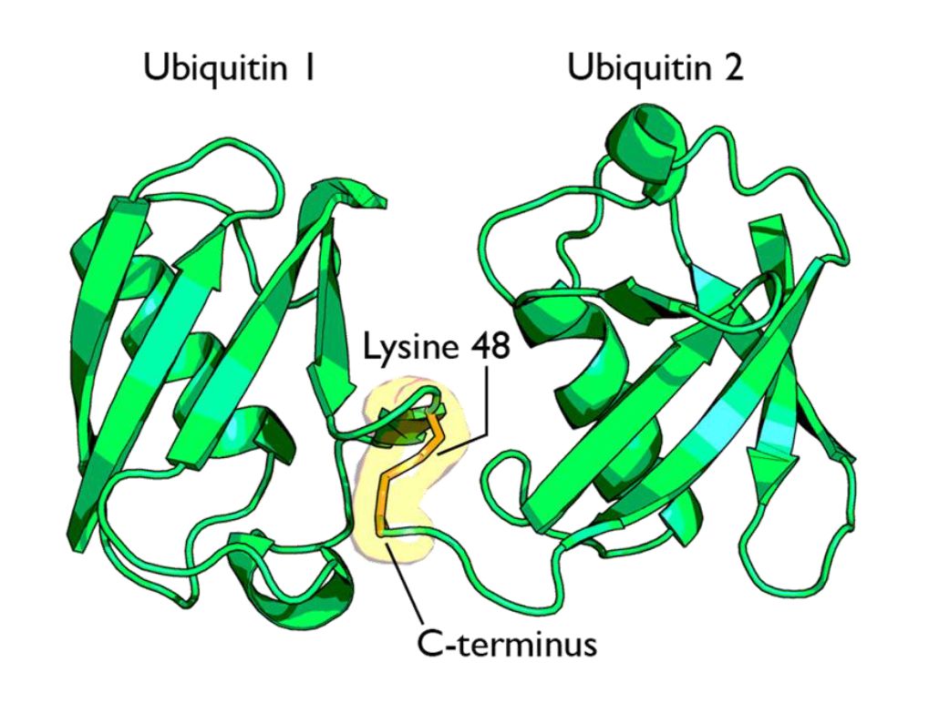
Fig 1 A cartoon representation of a lysine 48-linked diubiquitin molecule. The two ubiquitin chains are shown as green cartoons with each chain labelled. The components of the linkage are indicated and shown as orange sticks. By Rogerdodd (Own work) [CC BY-SA 3.0 (https://creativecommons.org/licenses/by-sa/3.0)], via Wikimedia Commons

The simplest type of chain is homogeneous (IOWs, ubiquitins are linked always at the same site). But many types of mixed and branched chains can also be found.

Let’s start with the most common situation: a poli-ubiquitination of (at least) 4 ubiqutins, linearly linked at K48. This is the common signal for proteasome degradation.

By the way, the 26S proteasome is another molecular machine of incredible complexity, made of more than 30 different proteins. However, its structure and function are not the object of this OP, and therefore I will not deal with them here.

The ubiquitin code is not completely understood, at present, but a few aspects have been well elucidated. Table 1 sums up the most important and well known modes:

Code

Meaning

Polyubiquitination (4 or more) with links at K48 or at K11 Proteasomal degradation
Monoubiqutination (single or multiple) Protein interactions, membrane trafficking, endocytosis
Polyubiquitination with links at K63 Endocytic trafficking, inflammation, translation, DNA repair.
Polyubiquitination with links at K63 (other links) Autophagic degradation of protein substrates
Polyubiquitination with links at K27, K29, K33 Non proteolytic processes
Rarer chain types (K6, K11) Under investigation


However, this is only a very partial approach. A recent bioinformatics paper:

An Interaction Landscape of Ubiquitin Signaling

(Paywall)

Has attempted for the first time a systematic approach to deciphering the whole code, using synthetic diubiquitins (all 8 possible variants) to identify the different interactors with those signals, and they identified, with two different methodologies,  111 and 53 selective interactors for linear polyUb chains, respectively. 46 of those interactors were identified by both methodologies.

The translation

But what “translates” the complex ubiquitin code, allowing ubiquinated proteins to met the right specific destiny? Again, we can refer to the diubiquitin paper quoted above.

How do cells decode this ubiquitin code into proper cellular responses? Recent studies have indicated that members of a protein family, ubiquitin-binding proteins (UBPs), mediate the recognition of ubiquitinated substrates. UBPs contain at least one of 20 ubiquitin-binding domains (UBDs) functioning as a signal adaptor to transmit the signal from ubiquitinated substrates to downstream effectors

But what are those “interactors” identified by the paper (at least 46 of them)? They are, indeed, complex proteins which recognize specific configurations of the “tag” (the ubiquitin chain), and link the tagged (ubiquinated) protein to other effector proteins which implement its final fate, or anyway contribute in deffrent forms to that final outcome.


The basic control of the procedure: the complexity of the ubiquitination process.

So, we have seen that ubiquitin chains work as tags, and that their coded signals are translated by specific interactors, so that the target protein may be linked to its final destiny, or contribute to the desired outcome. But we must still address one question: how is the ubiquitination of the different target proteins implemented? IOWs, what is the procedure that “writes” the specific codes associated to specific target proteins?

This is indeed the first step in the whole process. But it is also the most complex, and that’s why I have left it for the final part of the discussion.

Indeed, the ubiquitination process needs to realize the following aims:

  1. Identify the specific protein to be ubiquitinated
  2. Recognize the specific context in which that protein needs to be ubiquitinated
  3. Mark the target protein with the correct tag for the required fate or outcome

We have already seen that the ubiquitin system is involved in practically all different cellular paths and activities, and therefore we can expect that the implementation of the above functions must be a very complex thing.

And it is.

Now, we can certainly imagine that there are many different layers of regulation that may contribute to the general control of the procedure, specifically epigenetic levels, which are at present poorly understood. But there is one level that we can more easily explore and understand, and it is , as usual, the functional complexity of the proteins involved.

And, even at a first gross analysis, it is really easy to see that the functional complexity implied by this process is mind blowing.

Why? It is more than enough to consider the huge number of different proteins involved. Let’s see.

The ubiquitination process is well studied. It can be divided into three phases, each of which is implemented by a different kind of protein. The three steps, and the three kinds of proteins that implement them, take the name of E1, E2 and E3.
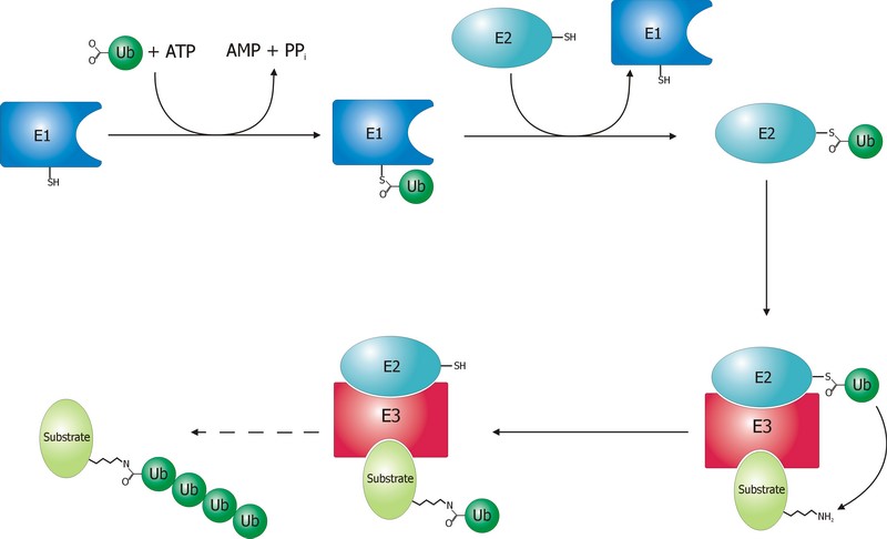
 

Fig. 2 Schematic diagram of the ubiquitylation system. Created by Roger B. Dodd: Rogerdodd at the English language Wikipedia [GFDL (http://www.gnu.org/copyleft/fdl.html) or CC-BY-SA-3.0 (http://creativecommons.org/licenses/by-sa/3.0/)], via Wikimedia Commons

 The E1 step of ubiquitination.

This is the first thing that happens, and it is also the simplest.

E1 is the process of activation of ubiquitin, and the E1 proteins is called E1 ubiquitin-activating enzyme. To put it simply, this enzyme “activates” the ubiquitin molecule in an ATP dependent process, preparing it for the following phases and attaching it to its active site cysteine residue. It is not really so simple, but for our purposes that can be enough.

This is a rather straightforward enzymatic reaction. In humans there are essentially two forms of E1 enzymes, UBA1 and UBA6, each of them about 1000 AAs long, and partially related at sequence level (42%).


The E2 step of ubiquitination.

The second step is ubiquitin conjugation. The activated ubiquitin is transferred from the E1 enzyme to the ubiquitin-conjugating enzyme, or E2 enzyme, where it is attached to a cysteine residue.

This apparently simple “transfer” is indeed a complex intermediate phase. Humans have about 40 different E2 molecules. The following paper:

E2 enzymes: more than just middle men

details some of the functional complexity existing at this level.

Abstract:

Ubiquitin-conjugating enzymes (E2s) are the central players in the trio of enzymes responsible for the attachment of ubiquitin (Ub) to cellular proteins. Humans have ∼40 E2s that are involved in the transfer of Ub or Ub-like (Ubl) proteins (e.g., SUMO and NEDD8). Although the majority of E2s are only twice the size of Ub, this remarkable family of enzymes performs a variety of functional roles. In this review, we summarize common functional and structural features that define unifying themes among E2s and highlight emerging concepts in the mechanism and regulation of E2s.

However, I will not go into details about these aspects, because we have better things to do: we still have to discuss the E3 phase!


The E3 step of ubiquitination.

This is the last phase of ubiquitination, where the ubiquitin tag is finally transferred to the target protein, as initial mono-ubiquitination, or to build an ubiquitin chain by following ubiqutination events. The proteins which implement this final passage are call E3 ubiquitin ligases. Here is the definition from Wikipedia:

A ubiquitin ligase (also called an E3 ubiquitin ligase) is a protein that recruits an E2 ubiquitin-conjugating enzyme that has been loaded with ubiquitin, recognizes a protein substrate, and assists or directly catalyzes the transfer of ubiquitin from the E2 to the protein substrate.

It is rather obvious that the role of the E3 protein is very important and delicate. Indeed it:

  1. Recognizes and links the E2-ubiquitin complex
  2. Recognizes and links some specific target protein
  3. Builds the appropriate tag for that protein (Monoubiquitination, mulptiple monoubiquitination, or poliubiquitination with the appropriate type of ubiquitin chain).
  4. And it does all those things at the right moment, in the right context, and for the right protein.

IOWs, the E3 protein writes the coded tag. It is, by all means, the central actor in our complex story.

So, here comes the really important point: how many different E3 ubiquitin ligases do we find in eukaryotic organisms? And the simple answer is: quite a lot!

Humans are supposed to have more than 600 different E3 ubiquitin ligases!

So, the human machinery for ubiquitination is about:

2 E1 proteins  –  40 E2 proteins – >600 E3 proteins

A real cascade of complexity!

OK, but even if we look at single celled eukaryotes we can already find an amazing level of complexity. In yeast, for example, we have:

1 or 2 E1 proteins  –  11 E2 proteins – 60-100 E3 proteins

See here:

The Ubiquitin–Proteasome System of Saccharomyces cerevisiae

Now, a very important point. Those 600+ E3 proteins that we find in humans are really different proteins. Of course, they have something in common: a specific domain.

From that point of view, they can be roughly classified in three groups according to the specific E3 domain:

  1. RING group: the RING finger domain ((Really Interesting New Gene) is a short domain of zinc finger type, usually 40 to 60 amino acids. This is the biggest group of E3s (about 600)
  2. HECT domain (homologous to the E6AP carboxyl terminus): this is a bigger domain (about 350 AAs). Located at the C terminus of the protein. It has a specific ligase activity, different from the RING   In humans we have approximately 30 proteins of this type.
  3. RBR domain (ring between ring fingers): this is a common domain (about 150 AAs) where two RING fingers are separated by a region called IBR, a cysteine-rich zinc finger. Only a subset of these proteins are E3 ligases, in humans we have about 12 of them.

See also here.

OK, so these proteins have one of these three domains in common, usually the RING domain. The function of the domain is specifically to interact with the E2-ubiquitin complex to implement the ligase activity. But the domain is only a part of the molecule, indeed a small part of it. E3 ligases are usually big proteins (hundreds, and up to thousands of AAs). Each of these proteins has a very specific non domain sequence, which is probably responsible for the most important part of the function: the recognition of the specific proteins that each E3 ligase processes.

This is a huge complexity, in terms of functional information at sequence level.

Our map of the ubiquinating system in humans could now be summarized as follows:

2 E1 proteins  –  40 E2 proteins – 600+ E3 proteins + thousands of specific substrates

IOWs, each of hundreds of different complex proteins recognizes its specific substrates, and marks them with a shared symbolic code based on uniquitin and its many possible chains. And the result of that process is that proteins are destined to degradation by the proteasome or other mechanisms, and that protein interactions and protein signaling are regulated and made possible, and that practically all cellular functions are allowed to flow correctly and smoothly.

Finally, here are two further compoments of the ubuquitination system, which I will barely mention, to avoid making this OP too long.

Ubiquitin like proteins (Ubl):

A number of ubiquitin like proteins add to the complexity of the system. Here is the abstract from a review:

The eukaryotic ubiquitin family encompasses nearly 20 proteins that are involved in the posttranslational modification of various macromolecules. The ubiquitin-like proteins (UBLs) that are part of this family adopt the β-grasp fold that is characteristic of its founding member ubiquitin (Ub). Although structurally related, UBLs regulate a strikingly diverse set of cellular processes, including nuclear transport, proteolysis, translation, autophagy, and antiviral pathways. New UBL substrates continue to be identified and further expand the functional diversity of UBL pathways in cellular homeostasis and physiology. Here, we review recent findings on such novel substrates, mechanisms, and functions of UBLs.

These proteins include SUMO, Nedd8, ISB15, and many others.

Deubiquitinating enzymes (DUBs):

The process of ubiquitination, complex as it already is, is additionally regulated by these enzymes which can cleave ubiquitin from proteins and other molecules. Doing so, they can reverse the effects of ubiquitination, creating a delicately balanced regulatory network. In humans there are nearly 100 DUB genes, which can be classified into two main classes: cysteine proteases and metalloproteases.


By the way, here is a beautiful animation of the basic working of the ubiquitin-proteasome system in degrading damaged proteins:


A summary:

So, let’s try a final graphic summary of the whole ubiquitin system in humans:
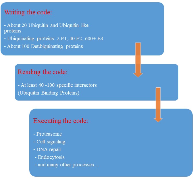
Fig 3 A graphic summary of the Ubiquitin System


Evolution of the Ubiquitin system?

The Ubiqutin system is essentially an eukaryotic tool. Of course, distant precursors for some of the main components have been “found” in prokaryotes. Here is the abstract from a paper that sums up what is known about the prokaryotic “origins” of the system:

Structure and evolution of ubiquitin and ubiquitin-related domains.

(Paywall)

Abstract:

Since its discovery over three decades ago, it has become abundantly clear that the ubiquitin (Ub) system is a quintessential feature of all aspects of eukaryotic biology. At the heart of the system lies the conjugation and deconjugation of Ub and Ub-like (Ubls) proteins to proteins or lipids drastically altering the biochemistry of the targeted molecules. In particular, it represents the primary mechanism by which protein stability is regulated in eukaryotes. Ub/Ubls are typified by the β-grasp fold (β-GF) that has additionally been recruited for a strikingly diverse range of biochemical functions. These include catalytic roles (e.g., NUDIX phosphohydrolases), scaffolding of iron-sulfur clusters, binding of RNA and other biomolecules such as co-factors, sulfur transfer in biosynthesis of diverse metabolites, and as mediators of key protein-protein interactions in practically every conceivable cellular context. In this chapter, we present a synthetic overview of the structure, evolution, and natural classification of Ub, Ubls, and other members of the β-GF. The β-GF appears to have differentiated into at least seven clades by the time of the last universal common ancestor of all extant organisms, encompassing much of the structural diversity observed in extant versions. The β-GF appears to have first emerged in the context of translation-related RNA-interactions and subsequently exploded to occupy various functional niches. Most biochemical diversification of the fold occurred in prokaryotes, with the eukaryotic phase of its evolution mainly marked by the expansion of the Ubl clade of the β-GF. Consequently, at least 70 distinct Ubl families are distributed across eukaryotes, of which nearly 20 families were already present in the eukaryotic common ancestor. These included multiple protein and one lipid conjugated forms and versions that functions as adapter domains in multimodule polypeptides. The early diversification of the Ubl families in eukaryotes played a major role in the emergence of characteristic eukaryotic cellular substructures and systems pertaining to nucleo-cytoplasmic compartmentalization, vesicular trafficking, lysosomal targeting, protein processing in the endoplasmic reticulum, and chromatin dynamics. Recent results from comparative genomics indicate that precursors of the eukaryotic Ub-system were already present in prokaryotes. The most basic versions are those combining an Ubl and an E1-like enzyme involved in metabolic pathways related to metallopterin, thiamine, cysteine, siderophore and perhaps modified base biosynthesis. Some of these versions also appear to have given rise to simple protein-tagging systems such as Sampylation in archaea and Urmylation in eukaryotes. However, other prokaryotic systems with Ubls of the YukD and other families, including one very close to Ub itself, developed additional elements that more closely resemble the eukaryotic state in possessing an E2, a RING-type E3, or both of these components. Additionally, prokaryotes have evolved conjugation systems that are independent of Ub ligases, such as the Pup system.


As usual, we are dealing here with distant similarities, but there is no doubt that the ubiquitin system as we know it appears in eukaryotes.

But what about its evolutionary history in eukaryotes?

We have already mentioned the extremely high conservation of ubiquitin itself.

UBA1, the main E1 enzyme, is rather well conserved from fungi to humans: 60% identity, 1282 bits, 1.21 bits per aminoacid (baa).

E2s are small enzymes, extremely conserved from fungi to humans: 86% identity, for example, for UB2D2, a 147 AAs molecule.

E3s, of course, are the most interesting issue. This big family of proteins behaves in different ways, consistently with its highly specific functions.

It is difficult to build a complete list of E3 proteins. I have downloaded from Uniprot a list of reviewed human proteins including “E3 ubiquitun ligase” in their name: a total of 223 proteins.

The mean evolutionary behavior of this group in metazoa is rather different from protein to protein. However, as a group these proteins exhibit an information jump in vertebrates which is significantly higher than the jump in all other proteins:
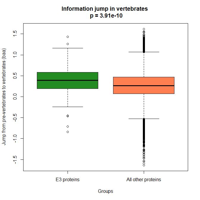
 

Fig. 4 Boxplots of the distribution of human conserved information jump from pre-vertebrates to vertebrates in 223 E3 ligase proteins and in all other human proteins. The difference is highly significant.


As we already know, this is evidence that this class of proteins is highly engineered in the transition to vertebrates. That is consistent with the need to finely regulate many cellular processes, most of which are certainly highly specific for different groups of organisms.

The highest vertebrate jump, in terms of bits per aminoacid, is shown in my group by the E3 ligase TRIM62. also known as DEAR1 (Q9BVG3), a 475 AAs long protein almost absent in pre-vertebrates (best hit 129 bits, 0.27 baa in Branchiostoma belcheri) and which flaunts an amazing jump of 1.433684 baa in cartilaginous fish (810 bits, 1.705263 baa).

But what is this protein? It is a master regulator tumor suppressor gene, implied in immunity, inflammation, tumor genesis.

See here:

TRIM Protein-Mediated Regulation of Inflammatory and Innate Immune Signaling and Its Association with Antiretroviral Activity

and here:

DEAR1 is a Chromosome 1p35 Tumor Suppressor and Master Regulator of TGFβ-Driven Epithelial-Mesenchymal Transition

This is just to show what a single E3 ligase can be involved in!

An opposite example, from the point of view of evolutionary history, is SIAH1, an E3 ligase implied in proteosomal degradation of proteins. It is a 282 AAs long protein, which already exhibits 1.787234 baa (504 bits) of homology in deuterostomes, indeed already 1.719858 baa in cnidaria. However, in fungi the best hit is only 50.8 bits (0.18 baa). So, this is a protein whose engineering takes place at the start of metazoa, and which exhibits only a minor further jump in vertebrates (0.29 baa), which brings the protein practically to its human form already in cartilaginous fish (280 identities out of 282, 99%). Practically a record.

So, we can see that E3 ligases are a good example of a class of proteins which perform different specific functions, and therefore exhibit different evolutionary histories: some, like TRIM62, are vertebrate quasi-novelties, others, like SIAH1, are metazoan quasi-novelties. And, of course, there are other behaviours, like for example BRCA1, Breast cancer type 1 susceptibility protein, a protein 1863 AAs long which only in mammals acquires part of its final sequence configuration in humans.

The following figure shows the evolutionary history of the three proteins mentioned above.
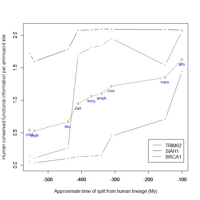
 

Fig. 5 Evolutionary history in metazoa of three E3 ligases (human conserved functional information)


An interesting example: NF-kB signaling

I will discuss briefly an example of how the Ubiquitin system interacts with some specific and complex final effector system. One of the best models for that is the NF-kB signaling.

NK-kB is a transcription factor family that is the final effector of a complex signaling pathway. I will rely mainly on the following recent free paper:

The Ubiquitination of NF-κB Subunits in the Control of Transcription

Here is the abstract:

Nuclear factor (NF)-κB has evolved as a latent, inducible family of transcription factors fundamental in the control of the inflammatory response. The transcription of hundreds of genes involved in inflammation and immune homeostasis require NF-κB, necessitating the need for its strict control. The inducible ubiquitination and proteasomal degradation of the cytoplasmic inhibitor of κB (IκB) proteins promotes the nuclear translocation and transcriptional activity of NF-κB. More recently, an additional role for ubiquitination in the regulation of NF-κB activity has been identified. In this case, the ubiquitination and degradation of the NF-κB subunits themselves plays a critical role in the termination of NF-κB activity and the associated transcriptional response. While there is still much to discover, a number of NF-κB ubiquitin ligases and deubiquitinases have now been identified which coordinate to regulate the NF-κB transcriptional response. This review will focus the regulation of NF-κB subunits by ubiquitination, the key regulatory components and their impact on NF-κB directed transcription.


The following figure sums up the main features of the canonical activation pathway:
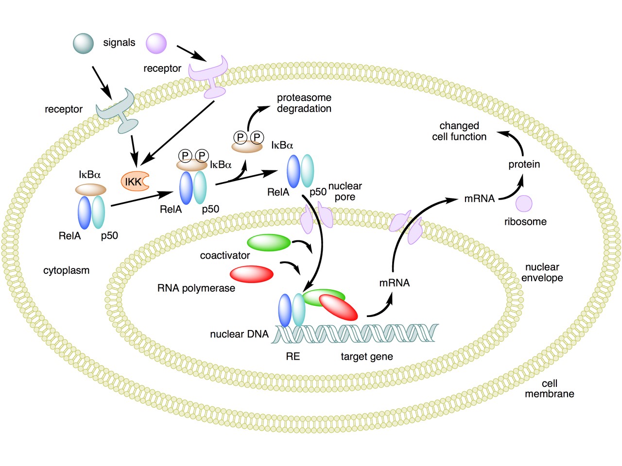
 

Fig. 6 A simple summary of the main steps in the canonical activayion pathway of NF-kB


Here the NF-κB TF is essentially the heterodimer RelA – p50. Before activation, the NF-κB (RelA – p50) dimer is kept in an inactive state and remains in the cytoplasm because it is linked to the IkB alpha protein, an inhibitor of its function.

Activation is mediated by a signal-receptor interaction, which starts the whole pathway. A lot of different signals can do that, adding to the complexity, but we will not discuss this part here.

As a consequence of receptor activation, another protein complex, IκB kinase (IKK), accomplishes the Phosphorylation of IκBα at serines 32 and 36. This is the signal for the ubiquitination of the IkB alpha inhibitor.

This ubiqutination targets IkB alpha for proteosomal degradation. But how is it achieved?

Well, things are not so simple. A whole protein complex is necessary, a complex which implements many different ubiquitinations in different contexts, including this one.

The complex is made by 3 basic proteins:

  • Cul1 (a scaffold protein, 776 AAs)
  • SKP1 (an adaptor protein, 163 AAs)
  • Rbx1 (a RING finger protein with E3 ligase activity, 108 AAs)

Plus:

  • An F-box protein (FBP) which changes in the different context, and confers specificity.

In our context, the F box protein is called beta TRC (605 AAs).
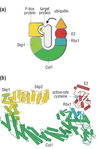
 

Fig. 7 A simple diagram of the SKP1 – beta TRC complex


Once the IkB alpha inhibitor is ubiquinated and degraded in the proteasome, the NF-κB dimer is free to translocate to the nucleus, and implement its function as a transcription factor (which is another complex issue, that we will not discuss).

OK, this is only the canonical activation of the pathway.

In the non canonical pathway (not shown in the figure) a different set of signals, receptors and activators acts on a different NF-κB dimer (RelB – p100). This dimer is not linked to any inhibitor, but is itself inactive in the cytoplasm. As a result of the signal, p100 is phosphorylated at serines 866 and 870. Again, this is the signal for ubiquitination.

This ubiquitination is performed by the same complex described above, but the result is different. P100 is only partially degraded in the proteasome, and is transformed into a smaller protein, p52, which remains linked to RelB. The RelB – p52 dimer is now an active NF-κB Transcription Factor, and it can relocate to the nucleus and act there.

But that’s not all.

  • You may remember that RelA (also called p 65) is one of the two components of NF-kB TF in the canonical pathway (the other being p 50). Well, RelA is heavily controlled by ubiquitination after it binds DNA in the nucleus to implement its TF activity. Ubiquitination (a very complex form of it) helps detachment of the TF from DNA, and its controlled degradation, avoiding sustained expression of NF-κB-dependent genes. For more details, see section 4 in the above quoted paper: “Ubiquitination of NF-κB”.
  • The activation of IKK in both the canonical and non canonical pathway after signal – receptor interaction is not so simple as depicted in Fig. 6. For more details, look at Fig. 1 in this paper: Ubiquitin Signaling in the NF-κB Pathway. You can see that, in the canonical pathway, the activation of IKK is mediated by many proteins, including TRAF2, TRAF6, TAK1, NEMO.
  • TRAF2 is a key regulator on many signaling pathways, including NF-kB. It is an E3 ubiquitin ligase. From Uniprot:  “Has E3 ubiquitin-protein ligase activity and promotes ‘Lys-63’-linked ubiquitination of target proteins, such as BIRC3, RIPK1 and TICAM1. Is an essential constituent of several E3 ubiquitin-protein ligase complexes, where it promotes the ubiquitination of target proteins by bringing them into contact with other E3 ubiquitin ligases.”
  • The same is true of TRAF6.
  • NEMO (NF-kappa-B essential modulator ) is also a key regulator. It is not an ubiquinating enzyme, but it is rather heavily regulated by ubiquitination. From Uniprot: “Regulatory subunit of the IKK core complex which phosphorylates inhibitors of NF-kappa-B thus leading to the dissociation of the inhibitor/NF-kappa-B complex and ultimately the degradation of the inhibitor. Its binding to scaffolding polyubiquitin seems to play a role in IKK activation by multiple signaling receptor pathways. However, the specific type of polyubiquitin recognized upon cell stimulation (either ‘Lys-63’-linked or linear polyubiquitin) and its functional importance is reported conflictingly.”
  • In the non canonical pathway, the activation of IKK alpha after signal – receptor interaction is mediated by other proteins, in particular one protein called NIK (see again Fig. 1 quoted above). Well, NIK is regulated by two different types of E3 ligases, with two different types of polyubiquitination:
    • cIAP E3 ligase inactivates it by constant degradation using a K48 chain
    • ZFP91 E3 ligase stabilizes it using a K63 chain

See here:

Non-canonical NF-κB signaling pathway.

In particular, Fig. 3

These are only some of the ways the ubiquitin system interacts with the very complex NF-kB signaling system. I hope that’s enough to show how two completely different and complex biological systems manage to cooperate by intricate multiple connections, and how the ubiquitin system can intervene at all levels of another process. What is true for the NF-kB signaling pathway is equally true for a lot of other biological systems, indeed for almost all basic cellular processes.

But this OP is already too long, and I have to stop here.

As usual, I want to close with a brief summary of the main points:

  1. The Ubiquitin system is a very important regulation network that shows two different signatures of design: amazing complexity and an articulated semiotic structure.
  2. The complexity is obvious at all levels of the network, but is especially amazing at the level of the hundreds of E3 ligases, that can recognize thousands of different substrates in different contexts.
  3. The semiosis is obvious in the Ubiquitin Code, a symbolic code of different ubiquitin configurations which serve as specific “tags” that point to different outcomes.
  4. The code is universally implemented and shared in eukaryotes, and allows control on almost all most important cellular processes.
  5. The code is written by the hundreds of E3 ligases. It is read by the many interactors with ubiquitin-binding domains (UBDs).
  6. The final outcome is of different types, including degradation, endocytosis, protein signaling, and so on.
  7. The interaction of the Ubiquitin System with other complex cellular pathways, like signaling pathways, is extremely complex and various, and happens at many different levels and by many different interacting proteins for each single pathway.

PS:

Thanks to DATCG for pointing to this video in three parts by Dr. Raymond Deshaies, was Professor of Biology at the California Institute of Technology and an Investigator of the Howard Hughes Medical Institute. On iBiology Youtube page:

A primer on the ubiquitin-proteasome system


Cullin-RING ubiquitin ligases: structure, structure, mechanism, and regulation


Targeting the ubiquitin-proteasome system in cancer

Comments
#42-44 UB, yes, I can work around it too. I found it a bit amusing to a degree. What he's advocating is screaming Design. The Codes, The Codes! ;-) It's why I posted them. I've not time to read Pattee, which I briefly looked at on your earlier post. Will look again when time permits. Pattee is right, your quote sums it up nicely.
"I am convinced that the problem of the origin of life cannot even be formulated without a better understanding of how molecules can function symbolically, that is, as records, codes, and signals. Or as I imply in my title, to understand origins, we need to know how a molecule becomes a message.
Indeed how is a correct code understood to be correct? After a billion, maybe trillion combinations? Hmmmmm. Where and when does time and resources become a factor? As more function in "junk" DNA is found, the limits to such random "success" becomes smaller as well. Will Dan Graur's threshold for "junk" DNA hold? Will he be found correct, or ENCODE correct and as Dan said, "evolution is wrong?" .DATCG
February 21, 2018
February
02
Feb
21
21
2018
08:32 PM
8
08
32
PM
PDT
#48. I really appreciate the numbers you've thought through and placed in your presentation. Understanding context is a real bonus for your readers.Upright BiPed
February 21, 2018
February
02
Feb
21
21
2018
07:11 PM
7
07
11
PM
PDT
Indeed, with about 1000 proteins involved specifically in the system, it is probably the cellular process with the greatest number of protein coding genes in the genome. We must remember that 1000 protein coding genes out of 20000 is almost 5% of the whole protein coding genome.
I am interested in how that compares numerically to the prokaryotic system.Upright BiPed
February 21, 2018
February
02
Feb
21
21
2018
07:00 PM
7
07
00
PM
PDT
On the other hand, the information coded by the ubiquitin code is written by the cell: it is essentially epigenetic information, not heritable genetic information. Of course, the writing is probably guided in some measure by the genome, and probably in some other measure by the epigenome, but the writing itself is implemented by the individual cell.
Fascinating isn't it. The cell is telling you what it is; layers of interdependence, and always yet another layer to be understood.Upright BiPed
February 21, 2018
February
02
Feb
21
21
2018
06:54 PM
6
06
54
PM
PDT
Just adding to our list of cell processes finely regulated by the Ubiquitin System. What are we missing? The central nervous system? Neurons? Synapses? Neuronal ubiquitin homeostasis. https://www.ncbi.nlm.nih.gov/pmc/articles/PMC3758786/
Abstract Neurons have highly specialized intracellular compartments that facilitate the development and activity of the nervous system. Ubiquitination is a post-translational modification that controls many aspects of neuronal function by regulating protein abundance. Disruption of this signaling pathway has been demonstrated in neurological disorders such as Parkinson’s disease, Amyotrophic Lateral Sclerosis and Angleman Syndrome. Since many neurological disorders exhibit ubiquitinated protein aggregates, the loss of neuronal ubiquitin homeostasis may be an important contributor of disease. This review discusses the mechanisms utilized by neurons to control the free pool of ubiquitin necessary for normal nervous system development and function as well as new roles of protein ubiquitination in regulating synaptic activity.
Following the de novo generation of ubiquitin in the cell body, ubiquitin is transported from the soma to distant locals like axons and dendrites. A single study in the literature indicates that ubiquitin is trafficked via slow axonal transport down the rat optic nerve [6]. This transport proceeds at a rate of approximately 3 mm/day, indicating that the length of time required for newly generated ubiquitin to reach synaptic terminals is on the order of days, or even weeks, in some neurons.
Since the original description of the ubiquitin proteasome system by Ciechanover, Hershko, and Rose in the late 1970’s and early 1980’s, numerous studies have linked ubiquitin to neuronal function [51]. While the contribution of protein ubiquitination in regulating synaptic protein abundance is well documented, there are hints in the literature of additional roles for ubiquitin at the synapse, which may be independent of protein degradation.
Nedd4 and Nedd4-2: ubiquitin ligases at work in the neuron.
Abstract Ubiquitination of proteins by the Nedd4 family of ubiquitin ligases is a significant mechanism in protein trafficking and degradation and provides for tight spatiotemporal regulation. Ubiquitination is gaining increasing recognition as a central mechanism underpinning the regulation of neuronal development and homeostasis in the brain. This review will focus on the Nedd4 and Nedd4-2 E3 ubiquitin ligases that are implicated in an increasing number of neuronal protein-protein interactions. Understanding of the contribution of Nedd4 and Nedd4-2 in regulating key functions in the brain is shedding new light on the ubiquitination signal not only in orchestrating degradation events but also in protein trafficking. Furthermore, the description of several novel Nedd4/4-2 targets in neurons is changing the way we conceptualize how neurons maintain normal function and how this is altered in disease.
Seven in Absentia E3 Ubiquitin Ligases: Central Regulators of Neural Cell Fate and Neuronal Polarity https://www.ncbi.nlm.nih.gov/pmc/articles/PMC5646344/
Abstract During neural development, neural precursors transition from a proliferative state within their germinal niches to a migratory state as they relocate to their final laminar positions. Transitions across these states are coupled with dynamic alterations in cellular polarity. This key feature can be seen throughout the developing vertebrate brain, in which neural stem cells give rise to multipolar or unpolarized transit-amplifying progenitors. These transit-amplifying progenitors then expand to give rise to mature neuronal lineages that become polarized as they initiate radial migration to their final laminar positions. The conventional understanding of the cellular polarity regulatory program has revolved around signaling cascades and transcriptional networks. In this review, we discuss recent discoveries concerning the role of the Siah2 ubiquitin ligase in initiating neuronal polarity during cerebellar development. Given the unique features of Siah ubiquitin ligases, we highlight some of the key substrates that play important roles in cellular polarity and propose a function for the Siah ubiquitin proteasome pathway in mediating a post-translational regulatory network to control the onset of polarization.
Spatial Organization of Ubiquitin Ligase Pathways Orchestrates Neuronal Connectivity https://www.ncbi.nlm.nih.gov/pmc/articles/PMC3622823/
Abstract Recent studies reveal that E3 ubiquitin ligases have essential functions in the establishment of neuronal circuits. Strikingly, a common emerging theme in these studies is that spatial organization of E3 ubiquitin ligases plays a critical role in the control of neuronal morphology and connectivity. E3 ubiquitin ligases localize to the nucleus, centrosome, Golgi apparatus, axon and dendrite cytoskeleton, and synapses in neurons. Localization of ubiquitin ligases within distinct subcellular compartments may facilitate neuronal responses to extrinsic cues as well as the ubiquitination of local substrates. Here, we will review the functions of neuronal E3 ubiquitin ligases at distinct subcellular locales and explore how they regulate neuronal morphology and function in the nervous system.
See in particular Fig. 2 and Fig. 3. And so on, and so on...gpuccio
February 21, 2018
February
02
Feb
21
21
2018
04:08 PM
4
04
08
PM
PDT
Upright Biped (and DATCG): And, finally: The complexity: While all the coding systems we have considered (and I would say all those quoted by DATCC at #41 from Barbieri’s autobiography page) are certainly hugely complex from any functional point of view, still the Ubiquitin System has a very special feature: the very high number of individual proteins involved. Indeed, with about 1000 proteins involved specifically in the system, it is probably the cellular process with the greatest number of protein coding genes in the genome. We must remember that 1000 protein coding genes out of 20000 is almost 5% of the whole protein coding genome. The only other example which comes to mind is olfactory receptors, which corrspond to about 1000 in the mammalian genome (but only 400 active in the human genome). But olfactory receptors are a class of not too long proteins which share a great sequence homology in many cases (50% or more), and so appear to be a diversification of similar molecules. Instead, E3 ligases, which make up the greatest part of proteins involved in the Ubiquitin System (600+), are a set of really distinct proteins, except for the shared domians. But, as already said, the shared domains are in general a minor part of the molecule. The RING domain, for example, which characterizes most of E3 ligases, is only 40 - 60 AAs long: in a class of proteins which has a mean length of about 500 AAs, that is certainly a minor part of the sequence information. The rest of the sequence is certainly involved in the specific function of each E3 ligase: to recognize the correct target protein, in the correct context. And each of the 600+ E3 ligases is completely different from each other in that respect. That makes a real lot of specific functional information in the system. Even from a proteomic point of view, the system represents a huge part of cell resources, its proteins amounting to about 1.3% of the total cell proteins, as stated here: The demographics of the ubiquitin system. http://www.cell.com/trends/cell-biology/fulltext/S0962-8924(15)00054-9 Many cell resources are used in many systems to generate variety. But while in some systems, like the immune system, variety is created using controlled variation on a limited number of genes, in the ubiquitin system (like in the olfactory system) variety is mostly paid for at genomic level. So, a lot of resources in the genome and the proteome are committed to that, an most of the sequence functional information is directly coded in the genome.gpuccio
February 21, 2018
February
02
Feb
21
21
2018
05:54 AM
5
05
54
AM
PDT
Upright Biped: Let's go on with the comparison between the Ubiquitin Code and the Genetic code. Reading the code: The genetic code, as we well know, is read by the 20 Aminoacyl tRNA synthetases, through the correctly charged tRNAs. This is where the code is really understood. The Ubiquitin code is similarly read by a number of specific proteins (maybe 40 - 100) which share a number of ubiquitin-binding domains (UBDs): about 20. So, the reading system seems to be a little more complex here, but essentially comparable to the reading system in the genetic code. The final effector: Here, too, the genetic code system seems to be relatively simpler: the final effector is the ribosome, and translation. However complex this is, it is still a rather homogeneous task. Not so in the Ubiquitin System: while there is one major effector (proteasomal degradation), we have seen that it is not the only one: indeed, a lot of important effects, in almost all cell systems, are mediated by specific effects on different protein - protein interactions and protein configurations, without involving proteasomal degradation in any way. More in next post.gpuccio
February 21, 2018
February
02
Feb
21
21
2018
05:24 AM
5
05
24
AM
PDT
Upright Biped: "sounds real familiar". And it is! There are many similarities with the best known example of semiotic system in biology: the genetic code. I would like to make a few reflections on the similarities and differences between the two systems: Nature of the code: The genetic code is in some measure more clear-cut. While redunndant, it is certainly strict and context independent. Codons have one unequivocal meaning. The ubiquitin code, like many other biologcial codes, seems to be nore fluid, and probably more context-dependent. In a sense, these seem to be higher forms of code, serving a more complex function. However, one specific feature of the ubiquitin code is that it relies practically on one single molecule, ubiquitin (and a few minor variants), and the main nature of the alphabet depends on the variety of chains that can be realized using the 8 internal switches. In that sense, there is an homogeneity of the tool which remind the genetic code, while other symbolic systems (like signal receptor systems) have greater variation and specificity in the nature of the signal. Writing the code: This is probably the gratest difference between the two systems. The information content coded by the genetic code is pre-existent. It is not written by the cell, it is simply inherited. In a sense, the information is written by the designer (ID point of view) or by the RV + NS process (neo-darwinist point of view). Indeed, one of the main objections against "third way speculations" is that there is no known mechanism in the cell that allows a flow of information from the phenotype to the genome (protein coding genes): IOWs, the cell does not know how to write new information (existing in the phenotype) at the level of protein coding genes. On the other hand, the information coded by the ubiquitin code is written by the cell: it is essentially epigenetic information, not heritable genetic information. Of course, the writing is probably guided in some measure by the genome, and probably in some other measure by the epigenome, but the writing itself is implemented by the individual cell. More in next post.gpuccio
February 21, 2018
February
02
Feb
21
21
2018
04:58 AM
4
04
58
AM
PDT
Upright BiPed (and DATCG): Welcome! :) :) It was really a beautiful surprise to awaken and find your many comments here! And your beautiful discussions with DATCG too... I was certain that you would like the semiosis angle. :) OK, our interlocutors, as usual, are nowhere to be seen, but at least I have some true friends! So thanks be given to you, DATCG, Dionisio. :) I am in a hurry now, so I will be back as soon as possible for some comments. I just wanted to thank all of you.gpuccio
February 21, 2018
February
02
Feb
21
21
2018
12:24 AM
12
12
24
AM
PDT
Pattee, 1969 How do we tell when there is communication in living systems? Most workers in the field probably do not worry too much about defining the idea of communication since so many concrete, experimental questions about developmental control do not depend on what communication means. But I am interested in the origin of life, and I am convinced that the problem of the origin of life cannot even be formulated without a better understanding of how molecules can function symbolically, that is, as records, codes, and signals. Or as I imply in my title, to understand origins, we need to know how a molecule becomes a message. Upright BiPed
February 20, 2018
February
02
Feb
20
20
2018
09:42 PM
9
09
42
PM
PDT
I think Gpuccio is on target re: semiosis and regulation.
No question about it.Upright BiPed
February 20, 2018
February
02
Feb
20
20
2018
09:33 PM
9
09
33
PM
PDT
Yes, Barbieri is an interesting fellow, and I surely appreciate his work. But there is a real problem -- setting aside for a moment the fact that he denies any room for design in biology. I can mentally work around that issue, but he also seams to find some difficulty is reconciling the encoding of biological information and the Peircean concept of interpretation. He sees them in some strange conflict with one another. I truly don't get his issue there. As far as I can see, Pattee put that issue completely to bed decades ago in the late 1960s. The actual physics of the system shot the deciding point. There is no such thing as a code without interpretation. - - - - - - - - anyway, not taking GP's excellent OP off courseUpright BiPed
February 20, 2018
February
02
Feb
20
20
2018
09:17 PM
9
09
17
PM
PDT
From Barbieri's autobiography page, interesting to note...
Many other organic codes have been discovered. Among them, the sequence codes (Trifonov 1987, 1989, 1999), the adhesive code (Redies and Takeichi 1996; Shapiro and Colman 1999), the splicing codes (Barbieri 2003; Fu 2004; Matlin et al. 2005; Pertea et al. 2007; Wang and Burge 2008; Barash et al. 2010; Dhir et al. 2010), the signal transduction codes (Barbieri 2003), the histone code (Strahl and Allis 2000; Jenuwein and Allis 2001; Turner 2000, 2002, 2007; Kühn and Hofmeyr 2014), the sugar code (Gabius 2000, 2009), the compartment codes (Barbieri 2003), the cytoskeleton codes (Barbieri 2003; Gimona 2008), the tubulin code (Verhey and Gaertig 2007), the nuclear signalling code(Maraldi 2008), the apoptosis code (Basañez and Hardwick 2008; Füllgrabe et al. 2010), the ubiquitin code (Komander and Rape 2012), the bioelectric code (Tseng and Levin 2013; Levin 2014), the glycomic code (Buckeridge and De Souza 2014) and the acoustic codes (Farina and Pieretti 2014).
How many other codes not included or to be found in future? Layer upon layer.
These discoveries have extraordinary consequences because they change our reconstruction and our understanding of the history of life. Any time that a new organic code came into being, something totally new appeared in Nature, something that had never existed before.
I think Gpuccio is on target re: semiosis and regulation. .DATCG
February 20, 2018
February
02
Feb
20
20
2018
08:48 PM
8
08
48
PM
PDT
#37 UB Arthur Hunt, one can hope. I thought he might provide some good insight on evolution of the Spliceosome via some of the comments he put forward. But nothing since then.DATCG
February 20, 2018
February
02
Feb
20
20
2018
08:40 PM
8
08
40
PM
PDT
Good to see you UB. Was responding on GPuccio's other point. #30 in his line below...
And the key word is: semiosis and regulation.
Yes, and look forward to hearing from UB on semiosis. ---------------------- :) ha! Great timing UB. I almost posted then saw your comments. So funny as I found your post form March 2016 just minutes earlier and was reading it and about Marcello Barbieri. Here is his autobiography... Marchello Barbieri Autobiography .DATCG
February 20, 2018
February
02
Feb
20
20
2018
08:33 PM
8
08
33
PM
PDT
Gpuccio... Thanks for your follow-up and answers. Quickly #30. I agree with it being a coordinated, parallel process. And I take note of your thoughts...
My personal idea is that eukaryotes and metazoa require, just from the beginning, a whole new design approach that is based on many interacting levels of regulation networks.
Agree with "require... whole new design approach" And new codes. Codes that must be interpreted, recognized and functional. Hardly amenable to random mutations. The amount of just-so happenstance for multiple proteins, signals and systems to simultaneously coordinate seem to be insurmountable.DATCG
February 20, 2018
February
02
Feb
20
20
2018
08:19 PM
8
08
19
PM
PDT
Perhaps Art Hunt will stop by and pick up on this excellent OP (and also get back to the unfinished business from your previous OP as well). :)Upright BiPed
February 20, 2018
February
02
Feb
20
20
2018
08:15 PM
8
08
15
PM
PDT
It reminds me of this... (if I may):
(from previous writing) Pattee recognized that representation and interpretation are two necessary and complementary roles in a very specific physical architecture; an organization where one arrangement of matter serves as a token of memory, while another arrangement of matter determines what is being represented by the arrangement of the token. Pattee further recognized that for this architecture to serve as the basis of open-ended variation, the referent would not be determined by the arrangement of the token. Instead, the referent would be independently specified by a second “interpreting” arrangement of matter, which he described as a non-integrable constraint.
Symbols do not exist in isolation but are part of a semiotic or linguistic system (Pattee, 1969a). Semiotic systems consist of (1) a discrete set of symbol structures (symbol vehicles) that can exist in a quiescent, rate-independent (non-dynamic) states, as in most memory storage, (2) a set of interpreting structures (non-integrable constraints, codes), and (3) an organism or system in which the symbols have a function (Pattee, 1986). – H.H. Pattee, "The Physics of Symbols: Bridging the Epistemic Cut". Biosystems. Vol. 60, pp. 5-21. 2001
In a semiotic system, a non-integrable constraint is an arrangement of matter that physically establishes a trajectory for the system (among alternatives) depending on the individual representation being translated. [...] They are referred to as “non-integrable” because a description of the constraints cannot be integrated with a lawful description of the system itself. Thus, the logician’s complementary roles of representation and interpretation are reflected in the physicist’s requirement of complementary descriptions as well -- one for the dynamic and another for the symbolic aspects of the system.
Upright BiPed
February 20, 2018
February
02
Feb
20
20
2018
08:05 PM
8
08
05
PM
PDT
Reading this:
But what “translates” the complex ubiquitin code, allowing ubiquinated proteins to met the right specific destiny? Again, we can refer to the diubiquitin paper quoted above.
How do cells decode this ubiquitin code into proper cellular responses? Recent studies have indicated that members of a protein family, ubiquitin-binding proteins (UBPs), mediate the recognition of ubiquitinated substrates. UBPs contain at least one of 20 ubiquitin-binding domains (UBDs) functioning as a signal adaptor to transmit the signal from ubiquitinated substrates to downstream effectors
But what are those “interactors” identified by the paper (at least 46 of them)? They are, indeed, complex proteins which recognize specific configurations of the “tag” (the ubiquitin chain), and link the tagged (ubiquinated) protein to other effector proteins which implement its final fate, or anyway contribute in deffrent forms to that final outcome.
....sounds real familiar. :) :)Upright BiPed
February 20, 2018
February
02
Feb
20
20
2018
08:02 PM
8
08
02
PM
PDT
GP, your presentation is just excellent. Where are your opponents, my friend? I don't see them. Are there no faithful left to carry the RV+NS banner into battle?Upright BiPed
February 20, 2018
February
02
Feb
20
20
2018
07:44 PM
7
07
44
PM
PDT
GP, you and I once spoken briefly about Marcello Barbieri holding his annual "Code Biology" conference in Italy. I suspect this is precisely the type of information covered in those labs and presentations.Upright BiPed
February 20, 2018
February
02
Feb
20
20
2018
07:38 PM
7
07
38
PM
PDT
Hello GP, I am just now seeing your fantastic OP. Man'o man, GP, what a wonderful job you've done in explaining the system and highlighting the scope of the issue. If you don't write a book, then there is something truly wrong with the world. I've just read the OP, haven't read the comments. It all functions via semiosis -- from the very start.Upright BiPed
February 20, 2018
February
02
Feb
20
20
2018
07:35 PM
7
07
35
PM
PDT
Dionisio: The bacteroides paper you link is really intriguing. It seems an obvious case or HGT with adaptation, but it is certainly atypical and stimulating. The most amazing fact is that this ubiquitin-like protein is used as a toxin against similar bacterial species. Infortunately, the mechanism of action of the protein in that role has not been clarified. that would really be interesting. It's interesting that the authors have no doubts that the bacterial protein was acquired, either from eukaryotes or from giant viruses. So, they opt for HGT just from the beginning. That seems obvious to me too, with the protein having 67% identities and 84% positives with human ubiquitin (bitscore 108 bits, E value 2e-38). It is clearly a derivation from eukaryotic ubiquitin. But I think a pure neo-darwinist could also propose some form of convergent evolution, or what? The simple truth is: 108 bits of information require an explanation. That's why the authors state (rightly): "The source from which ubb was acquired is not clear from existing genomic sequences." OK, but what about the thousands of bits that appear in thousand of individual protein, for example, at the origin of vertebrates? Or in eukaryotes? Don't they deserve some explanation too? Am I asking too much? :)gpuccio
February 20, 2018
February
02
Feb
20
20
2018
02:59 PM
2
02
59
PM
PDT
DATCG: Histone code, Ubiquitin code, cell signaling, and many other things, are essentially new layers of complexity that appear in eukaryotes, and become more stratified in metazoa. My personal idea is that eukaryotes and metazoa require, just from the beginning, a whole new design approach that is based on many interacting levels of regulation networks. And the key word is: semiosis and regulation. Of course, high levels of semiosis and regulation are already present in prokaryotes. But the eukaryotic world seems to be structured according to new concepts, and here is where we start to see these many parallel levels of control, each of them extremely important for practically all cell functions, each of them relatively independent, and yet each of them intertwined with all the others. So we have all the epigenetic layers of transcription regulation, which multiply the possibilities of a rather static genome: DNA methylation, histone code, chromatin remodeling, and so on. And we have the infinitely intricate netwotk of TFs, with their combinatorial working. And then the complex post-transcriptional regulation, the intron system, the spliceosome, miRNAs, lncRNAs. And a lot of different PTMs, including the Ubiquitination System which is the object of this discussion. And, last but not least, cell to cell signaling, and the multitude of signals and receptors and of transmission pathways that convey the symbolic meaning of the signal from the membrane receptor, through definite intemediate steps, and ultimately to the TF network and DNA. The amazing thing is that there is not one of these levels which does not control the same things as all the other levels, but in different ways, and that is not strictly interconnected with all the other levels. It's like a multiple control strategy whose complexity grows exponentially, and whose ultimate purpose can only be to attain functional outcomes which would be absolutely impossible without control, or with single level control. You ask: "So which code arrived 1st?" I think that the obsvious answer is: they are all part of the same project, they were conceived and implemented in a parallel process, just from the beginning, to realize a design of increasing complexity and efficiency of which we no equivalent is known.gpuccio
February 20, 2018
February
02
Feb
20
20
2018
02:45 PM
2
02
45
PM
PDT
DATCG: Thank you again for you very good thoughts. I would like to add some comments about the points you make, but I have not the time now. I will come back later! :)gpuccio
February 20, 2018
February
02
Feb
20
20
2018
09:11 AM
9
09
11
AM
PDT
Dionisio: Fig. 1 of the same paper is a very good summary of: "Writing, reading, and editing ubiquitin." Semiosis everywhere! Fig. 2 from the same paper is a very good summary of cullin based modular proteins, and it adds the even more complex APC/C structure (at least 14 different subunits!). From the Legend of the Figure:
Although the APC/C is closely related to CRLs and contains the cullin-homology subunit APC2 (Yu et al., 1998), it is structurally divergent. The APC/C is composed of at least 14 different subunits, including the RING subunit APC11, plus one of two coactivators (CDC20 and CDH1) that also participate in substrate binding (Sivakumar and Gorbsky, 2015). The APC/C operates in mitosis and G1 and is mostly known for its ability to degrade mitotic cyclins and other mitotic factors so that chromosomes are separated and mitotic exit ensues (Zhou et al., 2016).
gpuccio
February 20, 2018
February
02
Feb
20
20
2018
06:16 AM
6
06
16
AM
PDT
Dionisio: I have been missing your support! :) Yes, mitosis and cell division seem to be under the realm of ubiquitin control, too. About the paper you referenced at #22, it is rather surprising how some concept constantly recur in ubiquitin literature. For example, just from the abstract of this paper: dynamicity plasticity versatile fine-tune variety signals conceptual challenges Maybe my focus on semiosis in the OP is not so bad! :)gpuccio
February 20, 2018
February
02
Feb
20
20
2018
06:08 AM
6
06
08
AM
PDT
Gpuccio, Nice, glad videos are of help :) I've yet to get through the papers you and Dionisio have shared. Like at your #14 post... "So, this is another example of two complex symbolic codes (histone code and ubiquitin code) strongly interacting, in a highly dynamic and articulated pattern." So which code arrived 1st? Simultaneous Code building by random mutations and natural selection? Doubtful. Obviously Histone must be in place or can you imagine the journey along the length of DNA not packaged by Histones? And did ubiquitin just pop up one day and say, voila, I'm code and recognize histone binding targets and building pathways for DNA repair? Obviiously, I'm being a bit loose and humorous, but there's a point. And simple questions. If the two codes do not arise at same time or not built simultaneously working together, then what? So neo-Darwinist have addressed this, but how adequate is their explanation? What happens if ubiguitin code and enzyme regulation are not available? What happens to DNA Repair? And how often is repair required, double-stranded that is? Curious what neo-Darwinist propose as evolutionary history for multiple codes, pathways, signal recognition and repair functions rising together. As well as coordination of these systems. Will have more time to review tonight. Very good work Gpuccio! Thanks again :)DATCG
February 20, 2018
February
02
Feb
20
20
2018
04:10 AM
4
04
10
AM
PDT
Human gut Bacteroides species produce different types of toxins that antagonize closely related members of the gut microbiota. Some are toxic effectors delivered by type VI secretion systems, and others are non-contact-dependent secreted antimicrobial proteins. Many strains of Bacteroides fragilis secrete antimicrobial molecules, but only one of these toxins has been described to date (Bacteroidales secreted antimicrobial protein 1 [BSAP-1]). In this study, we describe a novel secreted protein produced by B. fragilis strain 638R that mediated intraspecies antagonism. Using transposon mutagenesis and deletion mutation, we identified a gene encoding a eukaryotic-like ubiquitin protein (BfUbb) necessary for toxin activity against a subset of B. fragilis strains. The addition of ubb into a heterologous background strain conferred toxic activity on that strain. We found this gene to be one of the most highly expressed in the B. fragilis genome. The mature protein is 84% similar to human ubiquitin but has an N-terminal signal peptidase I (SpI) signal sequence and is secreted extracellularly. We found that the mature 76-amino-acid synthetic protein has very potent activity, confirming that BfUbb mediates the activity. Analyses of human gut metagenomic data sets revealed that ubb is present in 12% of the metagenomes that have evidence of B. fragilis. As 638R produces both BSAP-1 and BfUbb, we performed a comprehensive analysis of the toxin activity of BSAP-1 and BfUbb against a set of 40 B. fragilis strains, revealing that 75% of B. fragilis strains are targeted by one or the other of these two secreted proteins of strain 638R.
Chatzidaki-Livanis, Maria & Coyne, Michael & G. Roelofs, Kevin & R. Gentyala, Rahul & M. Caldwell, Jarreth & E. Comstock, Laurie. (2017). Gut Symbiont Bacteroides fragilis Secretes a Eukaryotic-Like Ubiquitin Protein That Mediates Intraspecies Antagonism. mBio. 8. e01902-17. 10.1128/mBio.01902-17.
Dionisio
February 19, 2018
February
02
Feb
19
19
2018
07:53 PM
7
07
53
PM
PDT
Ubiquitination is a post-translational modification that defines the cellular fate of intracellular proteins. It can modify their stability, their activity, their subcellular location, and even their interacting pattern. This modification is a reversible event whose implementation is easy and fast. It contributes to the rapid adaptation of the cells to physiological intracellular variations and to intracellular or environmental stresses. E2F1 (E2 promoter binding factor 1) transcription factor is a potent cell cycle regulator. It displays contradictory functions able to regulate both cell proliferation and cell death. Its expression and activity are tightly regulated over the course of the cell cycle progression and in response to genotoxic stress. I discuss here the most recent evidence demonstrating the role of ubiquitination in E2F1’s regulation.
Dubrez, Laurence. (2017). Regulation of E2F1 Transcription Factor by Ubiquitin Conjugation. International Journal of Molecular Sciences. 18. 2188. 10.3390/ijms18102188.
Dionisio
February 19, 2018
February
02
Feb
19
19
2018
07:48 PM
7
07
48
PM
PDT
1 29 30 31 32

Leave a Reply