Uncommon Descent Serving The Intelligent Design Community

Cilia are more complex than thought

Share
Facebook
Twitter
LinkedIn
Flipboard
Print
Email
Scientists unravel mysteries of cells’ whiplike extensions

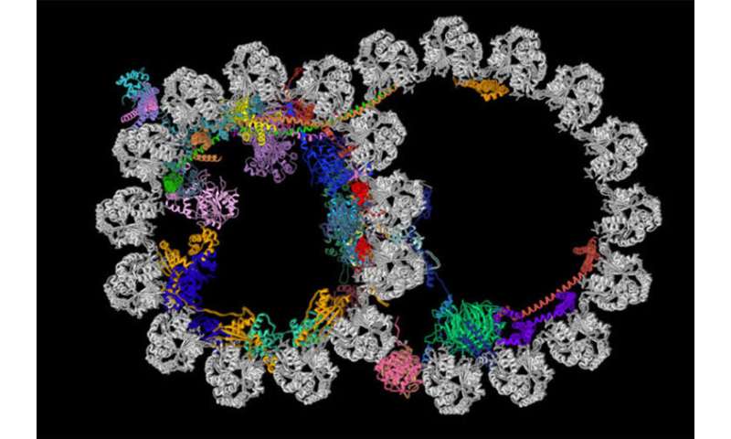
Cilia contain structures called ciliary doublet microtubules. Pictured is a cross section of one of these structures. A new study from the School of Medicine and Harvard Medical School has described the most detailed picture yet of these vital cellular structures. The microtubule is shown in gray, and the newly identified proteins decorating the microtubule are depicted in various colors. Credit: R. Zhang, A. Brown

Cilia (or flagella)—whiplike appendages on cells—do a lot of jobs that keep the body healthy:

“Before this work, everyone assumed these proteins inside cilia just stabilize the structure, which is true for a subset of the proteins, especially when you consider the forces produced by the continuous beating of the cilia,” Zhang said. “But based on how they are arranged inside this structure, we believe these proteins are doing many more things.”

Since many of the proteins protrude through the cilia, Zhang and his colleagues speculate that they may allow for communication between the inside and the outside of the ciliary microtubule doublets; govern the function of enzymes that make important biochemical reactions possible; and sense changes in the calcium concentration of the environment, which plays a role in triggering the cilia to beat.

Julia Evangelou Strait, “Scientists unravel mysteries of cells’ whiplike extensions” at Phys.Org

Follow UD News at Twitter!

:

Comments
The Paper's Summary:
The axoneme of motile cilia is the largest macromolecular machine of eukaryotic cells. In humans, impaired axoneme function causes a range of ciliopathies. Axoneme assembly, structure, and motility require a radially arranged set of doublet microtubules, each decorated in repeating patterns with non-tubulin components. We use single-particle cryo-electron microscopy to visualize and build an atomic model of the repeating structure of a native axonemal doublet microtubule, which reveals the identities, positions, repeat lengths, and interactions of 38 associated proteins, including 33 microtubule inner proteins (MIPs). The structure demonstrates how these proteins establish the unique architecture of doublet microtubules, maintain coherent periodicities along the axoneme, and stabilize the microtubules against the repeated mechanical stress induced by ciliary motility. Our work elucidates the architectural principles that underpin the assembly of this large, repetitive eukaryotic structure and provides a molecular basis for understanding the etiology of human ciliopathies.
PaV
November 24, 2019
November
11
Nov
24
24
2019
11:53 AM
11
11
53
AM
PDT
Let’s hope PavelU doesn’t post a nonsense comment making stupid claims.jawa
November 23, 2019
November
11
Nov
23
23
2019
03:04 PM
3
03
04
PM
PDT
This is a fascinating OP. Thanks.PeterA
November 23, 2019
November
11
Nov
23
23
2019
02:56 PM
2
02
56
PM
PDT

Leave a Reply