Uncommon Descent Serving The Intelligent Design Community

The Ubiquitin System: Functional Complexity and Semiosis joined together.

Share
Facebook
Twitter
LinkedIn
Flipboard
Print
Email

This is a very complex subject, so as usual I will try to stick to the essentials to make things as clear as possible, while details can be dealt with in the discussion.

It is difficult to define exactly the role of the Ubiquitin System. It is usually considered mainly a pathway which regulates protein degradation, but in reality its functions are much wider than that.

In essence, the US is a complex biological system which targets many different types of proteins for different final fates.

The most common “fate” is degradation of the protein. In that sense, the Ubiquitin System works together with another extremely complex cellular system, the proteasome. In brief, the Ubiquitin System “marks” proteins for degradation, and the proteasome degrades them.

It seems simple. It is not.

Ubiquitination is essentially one of many Post-Translational modifications (PTMs): modifications of proteins after their synthesis by the ribosome (translation). But, while most PTMs use simpler biochemical groups that are usually added to the target protein (for example, acetylation), in ubiquitination a whole protein (ubiquitin) is used as a modifier of the target protein.


The tool: Ubiquitin

Ubiquitin is a small protein (76 AAs). Its name derives from the simple fact that it  is found in most tissues of eukaryotic organisms.

Here is its aminoacid sequence:

MQIFVKTLTGKTITLEVEPSDTIENVKAKIQDKEGIPPD

QQRLIFAGKQLEDGRTLSDYNIQKESTLHLVLRLRGG

Essentially, it has two important properties:

  1. As said, it is ubiquitous in eukaryotes
  2. It is also extremely conserved in eukaryotes

In mammals, ubiquitin is not present as a single gene. It is encoded by 4 different genes: UBB, a poliubiquitin (3 Ub sequences); UBC, a poliubiquitin (9 Ub sequences); UBA52, a mixed gene (1   Ub sequence + the ribosomal protein L40); and RPS27A, again a mixed gene (1 Ub sequence + the ribosomal protein S27A). However, the basic ubiquitin sequence is always the same in all those genes.

Its conservation is one of the highest in eukaryotes. The human sequence shows, in single celled eukaryotes:

Naegleria: 96% conservation;  Alveolata: 100% conservation;  Cellular slime molds: 99% conservation; Green algae: 100% conservation; Fungi: best hit 100% conservation (96% in yeast).

Ubiquitin and Ubiquitin like proteins (see later) are characterized by a special fold, called  β-grasp fold.


The semiosis: the ubiquitin code

The title of this OP makes explicit reference to semiosis. Let’s try to see why.

The simplest way to say it is: ubiquitin is a tag. The addition of ubiquitin to a substrate protein marks that protein for specific fates, the most common being degradation by the proteasome.

But not only that. See, for example, the following review:

Nonproteolytic Functions of Ubiquitin in Cell Signaling

Abstract:

The small protein ubiquitin is a central regulator of a cell’s life and death. Ubiquitin is best known for targeting protein destruction by the 26S proteasome. In the past few years, however, nonproteolytic functions of ubiquitin have been uncovered at a rapid pace. These functions include membrane trafficking, protein kinase activation, DNA repair, and chromatin dynamics. A common mechanism underlying these functions is that ubiquitin, or polyubiquitin chains, serves as a signal to recruit proteins harboring ubiquitin-binding domains, thereby bringing together ubiquitinated proteins and ubiquitin receptors to execute specific biological functions. Recent advances in understanding ubiquitination in protein kinase activation and DNA repair are discussed to illustrate the nonproteolytic functions of ubiquitin in cell signaling.

Another important aspect is that ubiquitin is not one tag, but rather a collection of different tags. IOWs, a tag based code.

See, for example, here:

The Ubiquitin Code in the Ubiquitin-Proteasome System and Autophagy

(Paywall).

Abstract:

The conjugation of the 76 amino acid protein ubiquitin to other proteins can alter the metabolic stability or non-proteolytic functions of the substrate. Once attached to a substrate (monoubiquitination), ubiquitin can itself be ubiquitinated on any of its seven lysine (Lys) residues or its N-terminal methionine (Met1). A single ubiquitin polymer may contain mixed linkages and/or two or more branches. In addition, ubiquitin can be conjugated with ubiquitin-like modifiers such as SUMO or small molecules such as phosphate. The diverse ways to assemble ubiquitin chains provide countless means to modulate biological processes. We overview here the complexity of the ubiquitin code, with an emphasis on the emerging role of linkage-specific degradation signals (degrons) in the ubiquitin-proteasome system (UPS) and the autophagy-lysosome system (hereafter autophagy).

A good review of the basics of the ubiquitin code can be found here:

The Ubiquitin Code 

(Paywall)

It is particularly relevant, from an ID point of view, to quote the starting paragraph of that paper:

When in 1532 Spanish conquistadores set foot on the Inca Empire, they found a highly organized society that did not utilize a system of writing. Instead, the Incas recorded tax payments or mythology with quipus, devices in which pieces of thread were connected through specific knots. Although the quipus have not been fully deciphered, it is thought that the knots between threads encode most of the quipus’ content. Intriguingly, cells use a regulatory mechanism—ubiquitylation—that is reminiscent of quipus: During this reaction, proteins are modified with polymeric chains in which the linkage between ubiquitin molecules encodes information about the substrate’s fate in the cell.

Now, ubiquitin is usually linked to the target protein in chains. The first ubiquitin molecule is covalently bound through its C-terminal carboxylate group to a particular lysine, cysteine, serine, threonine or N-terminus of the target protein.

Then, additional ubiquitins are added to form a chain, and the C-terminus of the new ubiquitin is linked to one of seven lysine residues or the first methionine residue on the previously added ubiquitin.

IOWs, each ubiquitin molecule has seven lysine residues:

K6, K11, K27, K29, K33, K48, K63

And one N terminal methionine residue:

M1

And a new ubiquitin molecule can be added at each of those 8 sites in the previous ubiquitin molecule. IOWs, those 8 sites in the molecule are configurable switches that can be used to build ubiquitin chains.

Her are the 8 sites, in red, in the ubiquitin molecule:

MQIFVKTLTGKTITLEVEPSDTIENVKAKIQDKEGIPPD

QQRLIFAGKQLEDGRTLSDYNIQKESTLHLVLRLRGG

Fig 1 shows two ubiquitin molecules joined at K48.
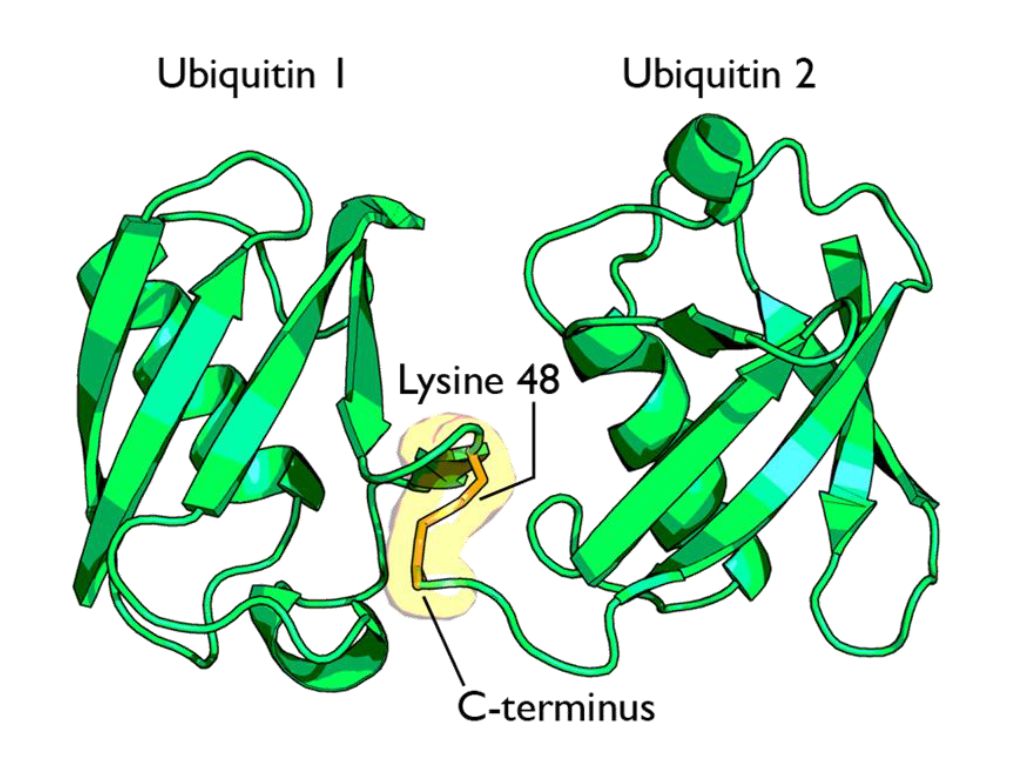
Fig 1 A cartoon representation of a lysine 48-linked diubiquitin molecule. The two ubiquitin chains are shown as green cartoons with each chain labelled. The components of the linkage are indicated and shown as orange sticks. By Rogerdodd (Own work) [CC BY-SA 3.0 (https://creativecommons.org/licenses/by-sa/3.0)], via Wikimedia Commons

The simplest type of chain is homogeneous (IOWs, ubiquitins are linked always at the same site). But many types of mixed and branched chains can also be found.

Let’s start with the most common situation: a poli-ubiquitination of (at least) 4 ubiqutins, linearly linked at K48. This is the common signal for proteasome degradation.

By the way, the 26S proteasome is another molecular machine of incredible complexity, made of more than 30 different proteins. However, its structure and function are not the object of this OP, and therefore I will not deal with them here.

The ubiquitin code is not completely understood, at present, but a few aspects have been well elucidated. Table 1 sums up the most important and well known modes:

Code

Meaning

Polyubiquitination (4 or more) with links at K48 or at K11 Proteasomal degradation
Monoubiqutination (single or multiple) Protein interactions, membrane trafficking, endocytosis
Polyubiquitination with links at K63 Endocytic trafficking, inflammation, translation, DNA repair.
Polyubiquitination with links at K63 (other links) Autophagic degradation of protein substrates
Polyubiquitination with links at K27, K29, K33 Non proteolytic processes
Rarer chain types (K6, K11) Under investigation


However, this is only a very partial approach. A recent bioinformatics paper:

An Interaction Landscape of Ubiquitin Signaling

(Paywall)

Has attempted for the first time a systematic approach to deciphering the whole code, using synthetic diubiquitins (all 8 possible variants) to identify the different interactors with those signals, and they identified, with two different methodologies,  111 and 53 selective interactors for linear polyUb chains, respectively. 46 of those interactors were identified by both methodologies.

The translation

But what “translates” the complex ubiquitin code, allowing ubiquinated proteins to met the right specific destiny? Again, we can refer to the diubiquitin paper quoted above.

How do cells decode this ubiquitin code into proper cellular responses? Recent studies have indicated that members of a protein family, ubiquitin-binding proteins (UBPs), mediate the recognition of ubiquitinated substrates. UBPs contain at least one of 20 ubiquitin-binding domains (UBDs) functioning as a signal adaptor to transmit the signal from ubiquitinated substrates to downstream effectors

But what are those “interactors” identified by the paper (at least 46 of them)? They are, indeed, complex proteins which recognize specific configurations of the “tag” (the ubiquitin chain), and link the tagged (ubiquinated) protein to other effector proteins which implement its final fate, or anyway contribute in deffrent forms to that final outcome.


The basic control of the procedure: the complexity of the ubiquitination process.

So, we have seen that ubiquitin chains work as tags, and that their coded signals are translated by specific interactors, so that the target protein may be linked to its final destiny, or contribute to the desired outcome. But we must still address one question: how is the ubiquitination of the different target proteins implemented? IOWs, what is the procedure that “writes” the specific codes associated to specific target proteins?

This is indeed the first step in the whole process. But it is also the most complex, and that’s why I have left it for the final part of the discussion.

Indeed, the ubiquitination process needs to realize the following aims:

  1. Identify the specific protein to be ubiquitinated
  2. Recognize the specific context in which that protein needs to be ubiquitinated
  3. Mark the target protein with the correct tag for the required fate or outcome

We have already seen that the ubiquitin system is involved in practically all different cellular paths and activities, and therefore we can expect that the implementation of the above functions must be a very complex thing.

And it is.

Now, we can certainly imagine that there are many different layers of regulation that may contribute to the general control of the procedure, specifically epigenetic levels, which are at present poorly understood. But there is one level that we can more easily explore and understand, and it is , as usual, the functional complexity of the proteins involved.

And, even at a first gross analysis, it is really easy to see that the functional complexity implied by this process is mind blowing.

Why? It is more than enough to consider the huge number of different proteins involved. Let’s see.

The ubiquitination process is well studied. It can be divided into three phases, each of which is implemented by a different kind of protein. The three steps, and the three kinds of proteins that implement them, take the name of E1, E2 and E3.
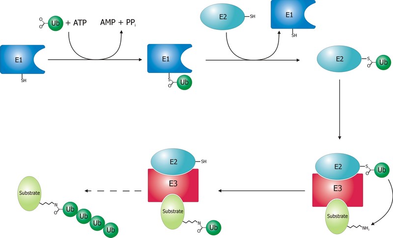
 

Fig. 2 Schematic diagram of the ubiquitylation system. Created by Roger B. Dodd: Rogerdodd at the English language Wikipedia [GFDL (http://www.gnu.org/copyleft/fdl.html) or CC-BY-SA-3.0 (http://creativecommons.org/licenses/by-sa/3.0/)], via Wikimedia Commons

 The E1 step of ubiquitination.

This is the first thing that happens, and it is also the simplest.

E1 is the process of activation of ubiquitin, and the E1 proteins is called E1 ubiquitin-activating enzyme. To put it simply, this enzyme “activates” the ubiquitin molecule in an ATP dependent process, preparing it for the following phases and attaching it to its active site cysteine residue. It is not really so simple, but for our purposes that can be enough.

This is a rather straightforward enzymatic reaction. In humans there are essentially two forms of E1 enzymes, UBA1 and UBA6, each of them about 1000 AAs long, and partially related at sequence level (42%).


The E2 step of ubiquitination.

The second step is ubiquitin conjugation. The activated ubiquitin is transferred from the E1 enzyme to the ubiquitin-conjugating enzyme, or E2 enzyme, where it is attached to a cysteine residue.

This apparently simple “transfer” is indeed a complex intermediate phase. Humans have about 40 different E2 molecules. The following paper:

E2 enzymes: more than just middle men

details some of the functional complexity existing at this level.

Abstract:

Ubiquitin-conjugating enzymes (E2s) are the central players in the trio of enzymes responsible for the attachment of ubiquitin (Ub) to cellular proteins. Humans have ∼40 E2s that are involved in the transfer of Ub or Ub-like (Ubl) proteins (e.g., SUMO and NEDD8). Although the majority of E2s are only twice the size of Ub, this remarkable family of enzymes performs a variety of functional roles. In this review, we summarize common functional and structural features that define unifying themes among E2s and highlight emerging concepts in the mechanism and regulation of E2s.

However, I will not go into details about these aspects, because we have better things to do: we still have to discuss the E3 phase!


The E3 step of ubiquitination.

This is the last phase of ubiquitination, where the ubiquitin tag is finally transferred to the target protein, as initial mono-ubiquitination, or to build an ubiquitin chain by following ubiqutination events. The proteins which implement this final passage are call E3 ubiquitin ligases. Here is the definition from Wikipedia:

A ubiquitin ligase (also called an E3 ubiquitin ligase) is a protein that recruits an E2 ubiquitin-conjugating enzyme that has been loaded with ubiquitin, recognizes a protein substrate, and assists or directly catalyzes the transfer of ubiquitin from the E2 to the protein substrate.

It is rather obvious that the role of the E3 protein is very important and delicate. Indeed it:

  1. Recognizes and links the E2-ubiquitin complex
  2. Recognizes and links some specific target protein
  3. Builds the appropriate tag for that protein (Monoubiquitination, mulptiple monoubiquitination, or poliubiquitination with the appropriate type of ubiquitin chain).
  4. And it does all those things at the right moment, in the right context, and for the right protein.

IOWs, the E3 protein writes the coded tag. It is, by all means, the central actor in our complex story.

So, here comes the really important point: how many different E3 ubiquitin ligases do we find in eukaryotic organisms? And the simple answer is: quite a lot!

Humans are supposed to have more than 600 different E3 ubiquitin ligases!

So, the human machinery for ubiquitination is about:

2 E1 proteins  –  40 E2 proteins – >600 E3 proteins

A real cascade of complexity!

OK, but even if we look at single celled eukaryotes we can already find an amazing level of complexity. In yeast, for example, we have:

1 or 2 E1 proteins  –  11 E2 proteins – 60-100 E3 proteins

See here:

The Ubiquitin–Proteasome System of Saccharomyces cerevisiae

Now, a very important point. Those 600+ E3 proteins that we find in humans are really different proteins. Of course, they have something in common: a specific domain.

From that point of view, they can be roughly classified in three groups according to the specific E3 domain:

  1. RING group: the RING finger domain ((Really Interesting New Gene) is a short domain of zinc finger type, usually 40 to 60 amino acids. This is the biggest group of E3s (about 600)
  2. HECT domain (homologous to the E6AP carboxyl terminus): this is a bigger domain (about 350 AAs). Located at the C terminus of the protein. It has a specific ligase activity, different from the RING   In humans we have approximately 30 proteins of this type.
  3. RBR domain (ring between ring fingers): this is a common domain (about 150 AAs) where two RING fingers are separated by a region called IBR, a cysteine-rich zinc finger. Only a subset of these proteins are E3 ligases, in humans we have about 12 of them.

See also here.

OK, so these proteins have one of these three domains in common, usually the RING domain. The function of the domain is specifically to interact with the E2-ubiquitin complex to implement the ligase activity. But the domain is only a part of the molecule, indeed a small part of it. E3 ligases are usually big proteins (hundreds, and up to thousands of AAs). Each of these proteins has a very specific non domain sequence, which is probably responsible for the most important part of the function: the recognition of the specific proteins that each E3 ligase processes.

This is a huge complexity, in terms of functional information at sequence level.

Our map of the ubiquinating system in humans could now be summarized as follows:

2 E1 proteins  –  40 E2 proteins – 600+ E3 proteins + thousands of specific substrates

IOWs, each of hundreds of different complex proteins recognizes its specific substrates, and marks them with a shared symbolic code based on uniquitin and its many possible chains. And the result of that process is that proteins are destined to degradation by the proteasome or other mechanisms, and that protein interactions and protein signaling are regulated and made possible, and that practically all cellular functions are allowed to flow correctly and smoothly.

Finally, here are two further compoments of the ubuquitination system, which I will barely mention, to avoid making this OP too long.

Ubiquitin like proteins (Ubl):

A number of ubiquitin like proteins add to the complexity of the system. Here is the abstract from a review:

The eukaryotic ubiquitin family encompasses nearly 20 proteins that are involved in the posttranslational modification of various macromolecules. The ubiquitin-like proteins (UBLs) that are part of this family adopt the β-grasp fold that is characteristic of its founding member ubiquitin (Ub). Although structurally related, UBLs regulate a strikingly diverse set of cellular processes, including nuclear transport, proteolysis, translation, autophagy, and antiviral pathways. New UBL substrates continue to be identified and further expand the functional diversity of UBL pathways in cellular homeostasis and physiology. Here, we review recent findings on such novel substrates, mechanisms, and functions of UBLs.

These proteins include SUMO, Nedd8, ISB15, and many others.

Deubiquitinating enzymes (DUBs):

The process of ubiquitination, complex as it already is, is additionally regulated by these enzymes which can cleave ubiquitin from proteins and other molecules. Doing so, they can reverse the effects of ubiquitination, creating a delicately balanced regulatory network. In humans there are nearly 100 DUB genes, which can be classified into two main classes: cysteine proteases and metalloproteases.


By the way, here is a beautiful animation of the basic working of the ubiquitin-proteasome system in degrading damaged proteins:


A summary:

So, let’s try a final graphic summary of the whole ubiquitin system in humans:
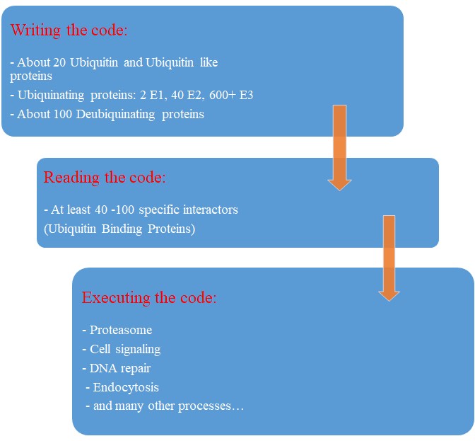
Fig 3 A graphic summary of the Ubiquitin System


Evolution of the Ubiquitin system?

The Ubiqutin system is essentially an eukaryotic tool. Of course, distant precursors for some of the main components have been “found” in prokaryotes. Here is the abstract from a paper that sums up what is known about the prokaryotic “origins” of the system:

Structure and evolution of ubiquitin and ubiquitin-related domains.

(Paywall)

Abstract:

Since its discovery over three decades ago, it has become abundantly clear that the ubiquitin (Ub) system is a quintessential feature of all aspects of eukaryotic biology. At the heart of the system lies the conjugation and deconjugation of Ub and Ub-like (Ubls) proteins to proteins or lipids drastically altering the biochemistry of the targeted molecules. In particular, it represents the primary mechanism by which protein stability is regulated in eukaryotes. Ub/Ubls are typified by the β-grasp fold (β-GF) that has additionally been recruited for a strikingly diverse range of biochemical functions. These include catalytic roles (e.g., NUDIX phosphohydrolases), scaffolding of iron-sulfur clusters, binding of RNA and other biomolecules such as co-factors, sulfur transfer in biosynthesis of diverse metabolites, and as mediators of key protein-protein interactions in practically every conceivable cellular context. In this chapter, we present a synthetic overview of the structure, evolution, and natural classification of Ub, Ubls, and other members of the β-GF. The β-GF appears to have differentiated into at least seven clades by the time of the last universal common ancestor of all extant organisms, encompassing much of the structural diversity observed in extant versions. The β-GF appears to have first emerged in the context of translation-related RNA-interactions and subsequently exploded to occupy various functional niches. Most biochemical diversification of the fold occurred in prokaryotes, with the eukaryotic phase of its evolution mainly marked by the expansion of the Ubl clade of the β-GF. Consequently, at least 70 distinct Ubl families are distributed across eukaryotes, of which nearly 20 families were already present in the eukaryotic common ancestor. These included multiple protein and one lipid conjugated forms and versions that functions as adapter domains in multimodule polypeptides. The early diversification of the Ubl families in eukaryotes played a major role in the emergence of characteristic eukaryotic cellular substructures and systems pertaining to nucleo-cytoplasmic compartmentalization, vesicular trafficking, lysosomal targeting, protein processing in the endoplasmic reticulum, and chromatin dynamics. Recent results from comparative genomics indicate that precursors of the eukaryotic Ub-system were already present in prokaryotes. The most basic versions are those combining an Ubl and an E1-like enzyme involved in metabolic pathways related to metallopterin, thiamine, cysteine, siderophore and perhaps modified base biosynthesis. Some of these versions also appear to have given rise to simple protein-tagging systems such as Sampylation in archaea and Urmylation in eukaryotes. However, other prokaryotic systems with Ubls of the YukD and other families, including one very close to Ub itself, developed additional elements that more closely resemble the eukaryotic state in possessing an E2, a RING-type E3, or both of these components. Additionally, prokaryotes have evolved conjugation systems that are independent of Ub ligases, such as the Pup system.


As usual, we are dealing here with distant similarities, but there is no doubt that the ubiquitin system as we know it appears in eukaryotes.

But what about its evolutionary history in eukaryotes?

We have already mentioned the extremely high conservation of ubiquitin itself.

UBA1, the main E1 enzyme, is rather well conserved from fungi to humans: 60% identity, 1282 bits, 1.21 bits per aminoacid (baa).

E2s are small enzymes, extremely conserved from fungi to humans: 86% identity, for example, for UB2D2, a 147 AAs molecule.

E3s, of course, are the most interesting issue. This big family of proteins behaves in different ways, consistently with its highly specific functions.

It is difficult to build a complete list of E3 proteins. I have downloaded from Uniprot a list of reviewed human proteins including “E3 ubiquitun ligase” in their name: a total of 223 proteins.

The mean evolutionary behavior of this group in metazoa is rather different from protein to protein. However, as a group these proteins exhibit an information jump in vertebrates which is significantly higher than the jump in all other proteins:
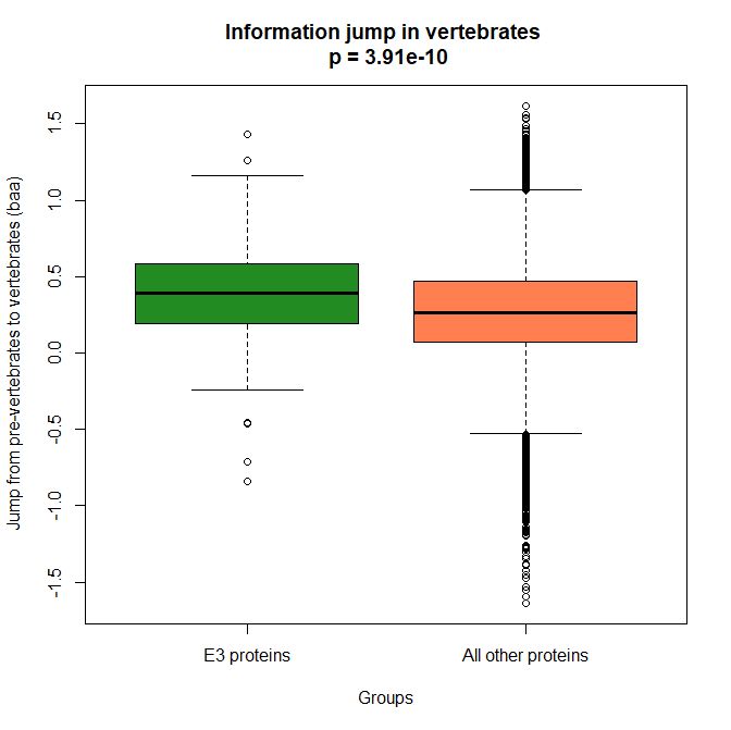
 

Fig. 4 Boxplots of the distribution of human conserved information jump from pre-vertebrates to vertebrates in 223 E3 ligase proteins and in all other human proteins. The difference is highly significant.


As we already know, this is evidence that this class of proteins is highly engineered in the transition to vertebrates. That is consistent with the need to finely regulate many cellular processes, most of which are certainly highly specific for different groups of organisms.

The highest vertebrate jump, in terms of bits per aminoacid, is shown in my group by the E3 ligase TRIM62. also known as DEAR1 (Q9BVG3), a 475 AAs long protein almost absent in pre-vertebrates (best hit 129 bits, 0.27 baa in Branchiostoma belcheri) and which flaunts an amazing jump of 1.433684 baa in cartilaginous fish (810 bits, 1.705263 baa).

But what is this protein? It is a master regulator tumor suppressor gene, implied in immunity, inflammation, tumor genesis.

See here:

TRIM Protein-Mediated Regulation of Inflammatory and Innate Immune Signaling and Its Association with Antiretroviral Activity

and here:

DEAR1 is a Chromosome 1p35 Tumor Suppressor and Master Regulator of TGFβ-Driven Epithelial-Mesenchymal Transition

This is just to show what a single E3 ligase can be involved in!

An opposite example, from the point of view of evolutionary history, is SIAH1, an E3 ligase implied in proteosomal degradation of proteins. It is a 282 AAs long protein, which already exhibits 1.787234 baa (504 bits) of homology in deuterostomes, indeed already 1.719858 baa in cnidaria. However, in fungi the best hit is only 50.8 bits (0.18 baa). So, this is a protein whose engineering takes place at the start of metazoa, and which exhibits only a minor further jump in vertebrates (0.29 baa), which brings the protein practically to its human form already in cartilaginous fish (280 identities out of 282, 99%). Practically a record.

So, we can see that E3 ligases are a good example of a class of proteins which perform different specific functions, and therefore exhibit different evolutionary histories: some, like TRIM62, are vertebrate quasi-novelties, others, like SIAH1, are metazoan quasi-novelties. And, of course, there are other behaviours, like for example BRCA1, Breast cancer type 1 susceptibility protein, a protein 1863 AAs long which only in mammals acquires part of its final sequence configuration in humans.

The following figure shows the evolutionary history of the three proteins mentioned above.
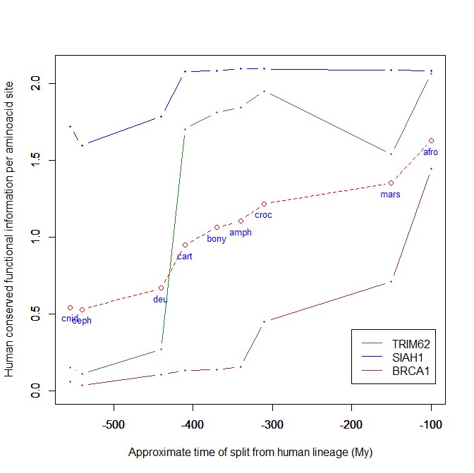
 

Fig. 5 Evolutionary history in metazoa of three E3 ligases (human conserved functional information)


An interesting example: NF-kB signaling

I will discuss briefly an example of how the Ubiquitin system interacts with some specific and complex final effector system. One of the best models for that is the NF-kB signaling.

NK-kB is a transcription factor family that is the final effector of a complex signaling pathway. I will rely mainly on the following recent free paper:

The Ubiquitination of NF-κB Subunits in the Control of Transcription

Here is the abstract:

Nuclear factor (NF)-κB has evolved as a latent, inducible family of transcription factors fundamental in the control of the inflammatory response. The transcription of hundreds of genes involved in inflammation and immune homeostasis require NF-κB, necessitating the need for its strict control. The inducible ubiquitination and proteasomal degradation of the cytoplasmic inhibitor of κB (IκB) proteins promotes the nuclear translocation and transcriptional activity of NF-κB. More recently, an additional role for ubiquitination in the regulation of NF-κB activity has been identified. In this case, the ubiquitination and degradation of the NF-κB subunits themselves plays a critical role in the termination of NF-κB activity and the associated transcriptional response. While there is still much to discover, a number of NF-κB ubiquitin ligases and deubiquitinases have now been identified which coordinate to regulate the NF-κB transcriptional response. This review will focus the regulation of NF-κB subunits by ubiquitination, the key regulatory components and their impact on NF-κB directed transcription.


The following figure sums up the main features of the canonical activation pathway:
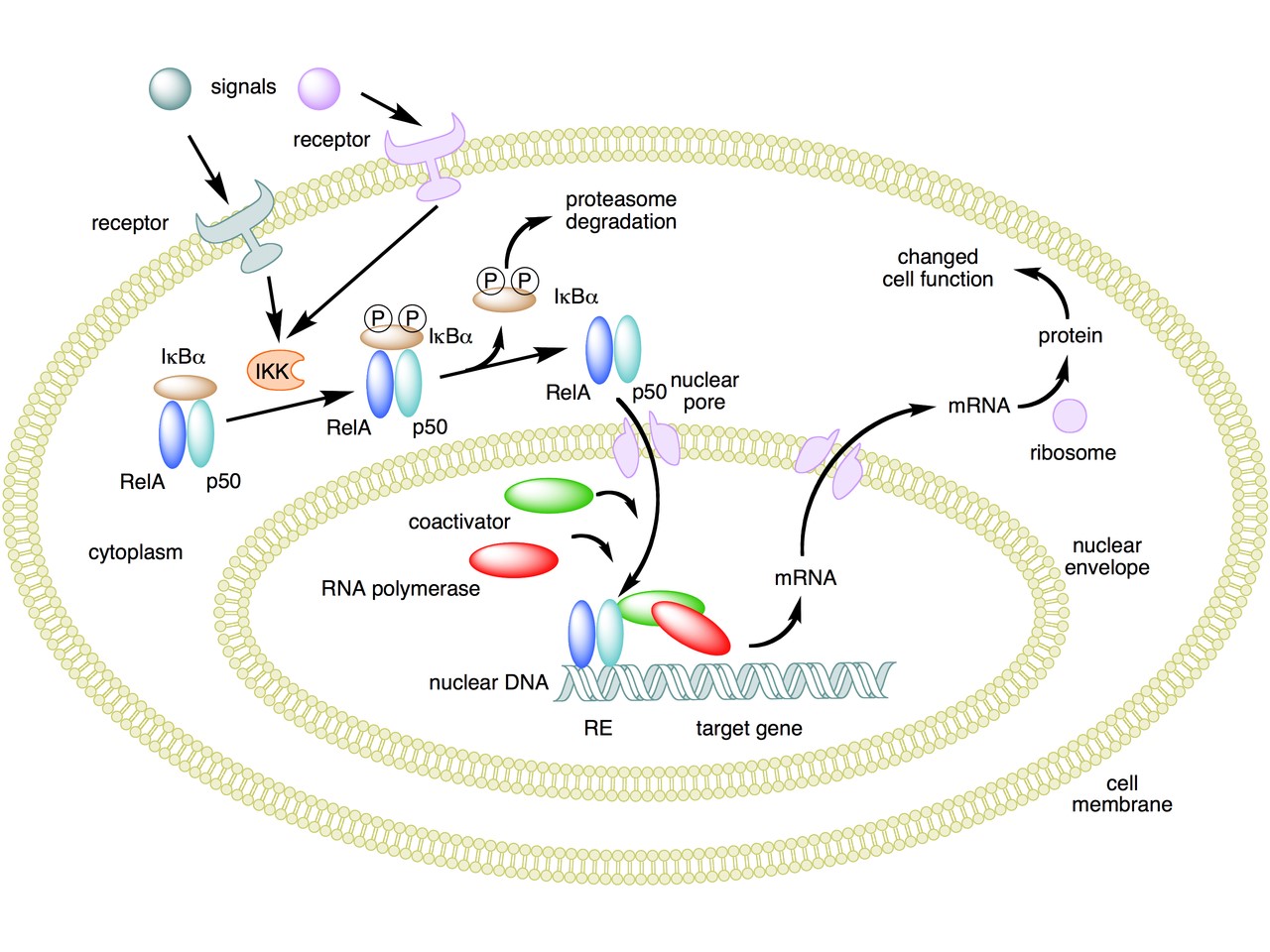
 

Fig. 6 A simple summary of the main steps in the canonical activayion pathway of NF-kB


Here the NF-κB TF is essentially the heterodimer RelA – p50. Before activation, the NF-κB (RelA – p50) dimer is kept in an inactive state and remains in the cytoplasm because it is linked to the IkB alpha protein, an inhibitor of its function.

Activation is mediated by a signal-receptor interaction, which starts the whole pathway. A lot of different signals can do that, adding to the complexity, but we will not discuss this part here.

As a consequence of receptor activation, another protein complex, IκB kinase (IKK), accomplishes the Phosphorylation of IκBα at serines 32 and 36. This is the signal for the ubiquitination of the IkB alpha inhibitor.

This ubiqutination targets IkB alpha for proteosomal degradation. But how is it achieved?

Well, things are not so simple. A whole protein complex is necessary, a complex which implements many different ubiquitinations in different contexts, including this one.

The complex is made by 3 basic proteins:

  • Cul1 (a scaffold protein, 776 AAs)
  • SKP1 (an adaptor protein, 163 AAs)
  • Rbx1 (a RING finger protein with E3 ligase activity, 108 AAs)

Plus:

  • An F-box protein (FBP) which changes in the different context, and confers specificity.

In our context, the F box protein is called beta TRC (605 AAs).
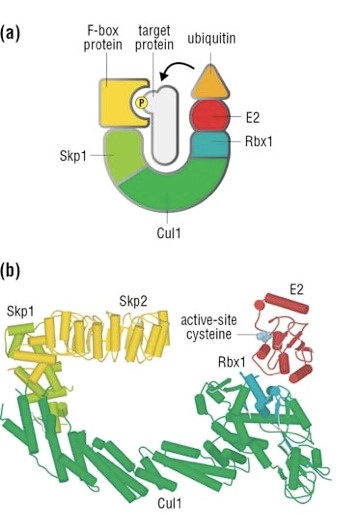
 

Fig. 7 A simple diagram of the SKP1 – beta TRC complex


Once the IkB alpha inhibitor is ubiquinated and degraded in the proteasome, the NF-κB dimer is free to translocate to the nucleus, and implement its function as a transcription factor (which is another complex issue, that we will not discuss).

OK, this is only the canonical activation of the pathway.

In the non canonical pathway (not shown in the figure) a different set of signals, receptors and activators acts on a different NF-κB dimer (RelB – p100). This dimer is not linked to any inhibitor, but is itself inactive in the cytoplasm. As a result of the signal, p100 is phosphorylated at serines 866 and 870. Again, this is the signal for ubiquitination.

This ubiquitination is performed by the same complex described above, but the result is different. P100 is only partially degraded in the proteasome, and is transformed into a smaller protein, p52, which remains linked to RelB. The RelB – p52 dimer is now an active NF-κB Transcription Factor, and it can relocate to the nucleus and act there.

But that’s not all.

  • You may remember that RelA (also called p 65) is one of the two components of NF-kB TF in the canonical pathway (the other being p 50). Well, RelA is heavily controlled by ubiquitination after it binds DNA in the nucleus to implement its TF activity. Ubiquitination (a very complex form of it) helps detachment of the TF from DNA, and its controlled degradation, avoiding sustained expression of NF-κB-dependent genes. For more details, see section 4 in the above quoted paper: “Ubiquitination of NF-κB”.
  • The activation of IKK in both the canonical and non canonical pathway after signal – receptor interaction is not so simple as depicted in Fig. 6. For more details, look at Fig. 1 in this paper: Ubiquitin Signaling in the NF-κB Pathway. You can see that, in the canonical pathway, the activation of IKK is mediated by many proteins, including TRAF2, TRAF6, TAK1, NEMO.
  • TRAF2 is a key regulator on many signaling pathways, including NF-kB. It is an E3 ubiquitin ligase. From Uniprot:  “Has E3 ubiquitin-protein ligase activity and promotes ‘Lys-63’-linked ubiquitination of target proteins, such as BIRC3, RIPK1 and TICAM1. Is an essential constituent of several E3 ubiquitin-protein ligase complexes, where it promotes the ubiquitination of target proteins by bringing them into contact with other E3 ubiquitin ligases.”
  • The same is true of TRAF6.
  • NEMO (NF-kappa-B essential modulator ) is also a key regulator. It is not an ubiquinating enzyme, but it is rather heavily regulated by ubiquitination. From Uniprot: “Regulatory subunit of the IKK core complex which phosphorylates inhibitors of NF-kappa-B thus leading to the dissociation of the inhibitor/NF-kappa-B complex and ultimately the degradation of the inhibitor. Its binding to scaffolding polyubiquitin seems to play a role in IKK activation by multiple signaling receptor pathways. However, the specific type of polyubiquitin recognized upon cell stimulation (either ‘Lys-63’-linked or linear polyubiquitin) and its functional importance is reported conflictingly.”
  • In the non canonical pathway, the activation of IKK alpha after signal – receptor interaction is mediated by other proteins, in particular one protein called NIK (see again Fig. 1 quoted above). Well, NIK is regulated by two different types of E3 ligases, with two different types of polyubiquitination:
    • cIAP E3 ligase inactivates it by constant degradation using a K48 chain
    • ZFP91 E3 ligase stabilizes it using a K63 chain

See here:

Non-canonical NF-κB signaling pathway.

In particular, Fig. 3

These are only some of the ways the ubiquitin system interacts with the very complex NF-kB signaling system. I hope that’s enough to show how two completely different and complex biological systems manage to cooperate by intricate multiple connections, and how the ubiquitin system can intervene at all levels of another process. What is true for the NF-kB signaling pathway is equally true for a lot of other biological systems, indeed for almost all basic cellular processes.

But this OP is already too long, and I have to stop here.

As usual, I want to close with a brief summary of the main points:

  1. The Ubiquitin system is a very important regulation network that shows two different signatures of design: amazing complexity and an articulated semiotic structure.
  2. The complexity is obvious at all levels of the network, but is especially amazing at the level of the hundreds of E3 ligases, that can recognize thousands of different substrates in different contexts.
  3. The semiosis is obvious in the Ubiquitin Code, a symbolic code of different ubiquitin configurations which serve as specific “tags” that point to different outcomes.
  4. The code is universally implemented and shared in eukaryotes, and allows control on almost all most important cellular processes.
  5. The code is written by the hundreds of E3 ligases. It is read by the many interactors with ubiquitin-binding domains (UBDs).
  6. The final outcome is of different types, including degradation, endocytosis, protein signaling, and so on.
  7. The interaction of the Ubiquitin System with other complex cellular pathways, like signaling pathways, is extremely complex and various, and happens at many different levels and by many different interacting proteins for each single pathway.

PS:

Thanks to DATCG for pointing to this video in three parts by Dr. Raymond Deshaies, was Professor of Biology at the California Institute of Technology and an Investigator of the Howard Hughes Medical Institute. On iBiology Youtube page:

A primer on the ubiquitin-proteasome system


Cullin-RING ubiquitin ligases: structure, structure, mechanism, and regulation


Targeting the ubiquitin-proteasome system in cancer

Comments
Hmm!Antonin
November 8, 2018
November
11
Nov
8
08
2018
06:09 AM
6
06
09
AM
PST
DATCG, OLV and all interested: Ubiquitin is definitely a friendly molecules. It even helps clarifying important issues. A recurring problem in our debates is that sometimes molecules that are extremely conserved in their evolutionary history are more tolerant than expected to sequence substitutions in the lab (for example, histones). This observation has been used many times by IDists and by neo-darwinists to state that sequence conservation could not be a good measure of functional constraint. I have always disagreed. As anyone who has read my OPs certainly knows, I am absolutely convinced that sequence conservation thorugh long evolutionary times is definitely a very good indicator of functional constraint. I have always argured that short term results in lab conditions are not a measure of lasting functional fitness, while sequence conservation definitely is. Now, this very interesting recent paper about ubiquitin seems to absolutely confirm my point: Extending chemical perturbations of the ubiquitin fitness landscape in a classroom setting reveals new constraints on sequence tolerance http://bio.biologists.org/content/7/7/bio036103.long
ABSTRACT: Although the primary protein sequence of ubiquitin (Ub) is extremely stable over evolutionary time, it is highly tolerant to mutation during selection experiments performed in the laboratory. We have proposed that this discrepancy results from the difference between fitness under laboratory culture conditions and the selective pressures in changing environments over evolutionary timescales. Building on our previous work (Mavor et al., 2016), we used deep mutational scanning to determine how twelve new chemicals (3-Amino-1,2,4-triazole, 5-fluorocytosine, Amphotericin B, CaCl2, Cerulenin, Cobalt Acetate, Menadione, Nickel Chloride, p-Fluorophenylalanine, Rapamycin, Tamoxifen, and Tunicamycin) reveal novel mutational sensitivities of ubiquitin residues. Collectively, our experiments have identified eight new sensitizing conditions for Lys63 and uncovered a sensitizing condition for every position in Ub except Ser57 and Gln62. By determining the ubiquitin fitness landscape under different chemical constraints, our work helps to resolve the inconsistencies between deep mutational scanning experiments and sequence conservation over evolutionary timescales.
gpuccio
July 27, 2018
July
07
Jul
27
27
2018
05:50 AM
5
05
50
AM
PST
OLV found an interesting paper! Cross-posting OLV's find here on Chromatin and Ubiquitin "crowbar" and Histone H2B... https://www.researchgate.net/publication/49765818_Chromatin_A_ubiquitin_crowbar_opens_chromatin
A ubiquitin crowbar opens chromatin Monoubiquitylation of histone H2B is found to disrupt condensation of chemically defined chromatin fibers. A novel fluorescence-based assay is used in concert with analytical ultracentrifugation to uncover the synergistic roles of histone acetylation and ubiquitylation on chromatin dynamics
Article in Nature Chemical Biology · February 2011 DOI: 10.1038/nchembio.514 · Source: PubMed Chromatin: A ubiquitin crowbar opens chromatin Originally posted by OLV in PaV's post on Chromatin... https://uncommondescent.com/intelligent-design/chromatin-topology-the-new-and-latest-functional-complexity/DATCG
July 25, 2018
July
07
Jul
25
25
2018
05:17 AM
5
05
17
AM
PST
Gpuccio @946, thanks and at #947... Ha! Evidence just keeps growing with nearly every discovery and paper! Design, not Blind! ;-) OLV @948-949, thanks as well! More reading to follow up on.DATCG
May 25, 2018
May
05
May
25
25
2018
08:02 PM
8
08
02
PM
PST
They mention the keyword "ubiquitin" here too: the function of the ubiquitin-like modifier DiSUMO-LIKE (DSUL) for early embryo development in maizeOLV
May 25, 2018
May
05
May
25
25
2018
05:45 PM
5
05
45
PM
PST
gpuccio: That's an interesting paper. Thanks. In 2018 they are still finding novel functions for the ubiquitin system components and also novel protein-protein interactions? BTW, do these discoveries help the case for neo-Darwinian macroevolution? https://www.molbiolcell.org/doi/pdf/10.1091/mbc.E17-04-0248
We demonstrate a novel function for the E3 Ub ligase UBR5 in regulation of ciliogenesis via maintenance of centriolar satellite stability. We also demonstrate a novel protein-protein interaction between UBR5 and the CSPP-L isoform of CSPP1, predominantly at the centrosome and surrounding centriolar satellites.
In summary, we have demonstrated a highly novel role for the E3 Ub ligase UBR5 in primary cilia maintenance/formation, with potential implications for understanding the molecular basis of key signalling pathways in development and disease.
OLV
May 10, 2018
May
05
May
10
10
2018
06:40 PM
6
06
40
PM
PST
To all: This is brand new: The E3 ubiquitin ligase UBR5 regulates centriolar satellite stability and primary cilia. https://www.ncbi.nlm.nih.gov/pubmed/29742019
Abstract: Primary cilia are crucial for signal transduction in a variety of pathways, including Hedgehog and Wnt. Disruption of primary cilia formation (ciliogenesis) is linked to numerous developmental disorders (known as ciliopathies) and diseases, including cancer. The Ubiquitin-Proteasome System (UPS) component UBR5 was previously identified as a putative positive regulator of ciliogenesis in a functional genomics screen. UBR5 is an E3 Ubiquitin ligase that is frequently deregulated in tumours, but its biological role in cancer is largely uncharacterised, partly due to a lack of understanding of interacting proteins and pathways. We validated the effect of UBR5 depletion on primary cilia formation using a robust model of ciliogenesis, and identified CSPP1, a centrosomal and ciliary protein required for cilia formation, as a UBR5-interacting protein. We show that UBR5 ubiquitylates CSPP1, and that UBR5 is required for cytoplasmic organization of CSPP1-comprising centriolar satellites in centrosomal periphery, suggesting that UBR5 mediated ubiquitylation of CSPP1 or associated centriolar satellite constituents is one underlying requirement for cilia expression. Hence, we have established a key role for UBR5 in ciliogenesis that may have important implications in understanding cancer pathophysiology.
Now, UBR5 is an E3 ligase of exceptional length: 2799 AAs. This is the "function" section at Uniprot: "E3 ubiquitin-protein ligase which is a component of the N-end rule pathway. Recognizes and binds to proteins bearing specific N-terminal residues that are destabilizing according to the N-end rule, leading to their ubiquitination and subsequent degradation (By similarity). Involved in maturation and/or transcriptional regulation of mRNA by activating CDK9 by polyubiquitination. May play a role in control of cell cycle progression. May have tumor suppressor function. Regulates DNA topoisomerase II binding protein (TopBP1) in the DNA damage response. Plays an essential role in extraembryonic development. Ubiquitinates acetylated PCK1. Also acts as a regulator of DNA damage response by acting as a suppressor of RNF168, an E3 ubiquitin-protein ligase that promotes accumulation of 'Lys-63'-linked histone H2A and H2AX at DNA damage sites, thereby acting as a guard against excessive spreading of ubiquitinated chromatin at damaged chromosomes." Now, this protein has an amazing jump in human-conserved information from pre-vertebrates to vertebrates: 2098 bits (0.75 baa) The human protein and the protein in cartilaginous fish (callorhincus milii) show the following homology: 4913 bits 2574 identities (92%) 2690 positives (95%) IOWs, this very long protein has remained almost the same for 400+ million years! Uniprot recognizes only two domains in the C terminal part: PABC (78 AAs) HECT (338 AAs) The Blast page recognizes the same two domains, plus one small putative zinc finger (67 AA) in the middle of the sequence, and an even smaller CUE domain (64 AAs) in the N terminal part. IOWs, more than 2200 AAs that make up the protein, and that are extremely conserved, do not correspond to known domains. This is certainly an amazing example of a highly specific and very long sequence, whose complex regulatory functions we can only barely imagine, and that exhibits almost 5000 bits of functional information (conserved from cartilaginous fish to humans) more than 2000 of them appearing for the first time in the transition to vertebrates.gpuccio
May 10, 2018
May
05
May
10
10
2018
04:59 PM
4
04
59
PM
PST
DATCG at #944: Thank you for cross-posting. Strange that blind evolution can so easily find targets that we cannot even predict.gpuccio
May 6, 2018
May
05
May
6
06
2018
10:54 AM
10
10
54
AM
PST
To all: This is an in depth analysis of the ERAD (ER-associated protein degradation) retrotransposition mechanism, already quoted in this discussion. Mechanistic insights into ER-associated protein degradation https://www.sciencedirect.com/science/article/pii/S0955067418300048
Abstract: Misfolded proteins of the endoplasmic reticulum (ER) are discarded by a conserved process, called ER-associated protein degradation (ERAD). ERAD substrates are retro-translocated into the cytosol, polyubiquitinated, extracted from the ER membrane, and ultimately degraded by the proteasome. Recent in vitro experiments with purified components have given insight into the mechanism of ERAD. ERAD substrates with misfolded luminal or intramembrane domains are moved across the ER membrane through a channel formed by the multispanning ubiquitin ligase Hrd1. Following polyubiquitination, substrates are extracted from the membrane by the Cdc48/p97 ATPase complex and transferred to the proteasome. We discuss the molecular mechanism of these processes and point out remaining open questions.
Unfortunately, the paper is paywalled. The general idewa is that proteins from the ER must be degraded in the cytosol by the proteasome. For that to happen, they must be "retrotranslocated" to be ubiquinated. It seems that the protein manily responsible for that is Hrd1, a transmembrane E3 ligase, which "exposes" the target protein to the cytosol and ubiquinates it. Another protein complex (Cdc48/p97 ATPase) then extracts the ubiquinated protein from the ER membrane, so that it can be degraded in the cytosol by the proteasome. The ERAD pathway exists in three different forms: ERAD-L, ERAD-M and ERAD-C. Each of them is implemented by different specific protein complexes. For ERAD-L (the pathway involved in degradation of intraluminal ER proteins) we have: a) A complex linked to the ER membrane, made of 4 different proteins: Hrd3, Usa1, Der1 and Hrd1, where both Hrd3 and Hrd1 are E3 ligases, and Hrd1 is the "channel" through which the target protein is retrotranslocated, exposed to the cytosol and ubquinated. b) A luminal protein, Yos9 c) Other ubiquinating components on the cytosol margin of the ER membrane, including Ubc7 (an E2 enzyme) and Cue1 d) A protein complex which extracts the ubiquinated target protein from the membrane, and releases it for proteosomal degradation in the cytosol, made of our well known Cdc48/VCP/p97 (or however you want to call it), Ufd1 (Ubiquitin recognition factor) and Npl4, plus at least one DUB (Otu1), but probably more than one. e) A couple of "shuttling factors", Rad23 and Dsk2, which each have both ubiquitin- and proteasome-binding domains, and which are probably responsible for transferring the target protein to the proteasome. Rather simple, isn't it? And this is only the ERAD-L pathway! :)gpuccio
May 6, 2018
May
05
May
6
06
2018
10:51 AM
10
10
51
AM
PST
Gpuccio @942, thanks :) I'm cross-posting your link from Defending Design OP here for the Ubiqitin Proteaome System: Predictive hypotheses are ineffectual in resolving complex biochemical systems. Abstract:
Scientific hypotheses may either predict particular unknown facts or accommodate previously-known data. Although affirmed predictions are intuitively more rewarding than accommodations of established facts, opinions divide whether predictive hypotheses are also epistemically superior to accommodation hypotheses. This paper examines the contribution of predictive hypotheses to discoveries of several bio-molecular systems. Having all the necessary elements of the system known beforehand, an abstract predictive hypothesis of semiconservative mode of DNA replication was successfully affirmed. However, in defining the genetic code whose biochemical basis was unclear, hypotheses were only partially effective and supplementary experimentation was required for its conclusive definition. Markedly, hypotheses were entirely inept in predicting workings of complex systems that included unknown elements. Thus, hypotheses did not predict the existence and function of mRNA, the multiple unidentified components of the protein biosynthesis machinery, or the manifold unknown constituents of the ubiquitin-proteasome system of protein breakdown. Consequently, because of their inability to envision unknown entities, predictive hypotheses did not contribute to the elucidation of of complex systems. As data-based accommodation theories remained the sole instrument to explain complex bio-molecular systems, the philosophical question of alleged advantage of predictive over accommodative hypotheses became inconsequential.
Imagine that, a blind belief in gradualist blind events cannot envision or predict functionally complex systems? That's an honest assessment at least and well founded. Yet Darwinist will claim Design Theorist lack imagination. And still cling to JUNK DNA as their savior. Yep, they missed out on the UPS as this well done OP and growing aggregated list of Ubiquitin Systems network topology and functionality keeps increasing day by day.DATCG
May 3, 2018
May
05
May
3
03
2018
10:52 AM
10
10
52
AM
PST
DATCG and all: This is from today's search: Neuronal Proteomic Analysis of the Ubiquitinated Substrates of the Disease-Linked E3 Ligases Parkin and Ube3a https://www.hindawi.com/journals/bmri/2018/3180413/
Abstract: Both Parkin and UBE3A are E3 ubiquitin ligases whose mutations result in severe brain dysfunction. Several of their substrates have been identified using cell culture models in combination with proteasome inhibitors, but not in more physiological settings. We recently developed the strategy to isolate ubiquitinated proteins in flies and have now identified by mass spectrometry analysis the neuronal proteins differentially ubiquitinated by those ligases. This is an example of how flies can be used to provide biological material in order to reveal steady state substrates of disease causing genes. Collectively our results provide new leads to the possible physiological functions of the activity of those two disease causing E3 ligases. Particularly, in the case of Parkin the novelty of our data originates from the experimental setup, which is not overtly biased by acute mitochondrial depolarisation. In the case of UBE3A, it is the first time that a nonbiased screen for its neuronal substrates has been reported.
Public access. Here are a few interesting remarks from the paper:
Both Parkin (PARK2) and UBE3A are E3 ubiquitin ligases for which mutations result in severe brain dysfunction, Familial Parkinson’s Disease (PD), and Angelman Syndrome (AS). In order to unravel the molecular mechanisms leading to these neurological dysfunctions it is necessary to identify and understand the role of their ubiquitinated substrates. --- However, even when proteins are correctly folded and functionally active in their final compartment, various factors can destabilise the proteins and irreversibly impair them. For this purpose, cells possess quality control mechanisms such as the Ubiquitin-Proteasome System (UPS) and autophagy that specifically degrade damaged proteins and organelles. --- Interestingly, ubiquitination is also involved in the regulation of autophagy [14–19]. In addition to its other roles, therefore, it is clear that ubiquitination serves as universal tag for substrate degradation, as all intracellular degradation pathways appear to be interconnected and governed by it. --- On our first application of this method, our group detected 121 ubiquitinated proteins in Drosophila neurons during embryonic development [149], including several key proteins involved in synaptogenesis and hence suggesting that UPS is important for proper neuronal arrangement. We later compared the ubiquitin landscape between developing and mature neurons in Drosophila melanogaster and identified 234 and 369 ubiquitinated proteins, respectively [154], some of which were found in both developmental stages. More interestingly, certain proteins are preferentially ubiquitinated in specific cell types during specific periods of the Drosophila life cycle, reinforcing the importance of using the appropriate cell type when studying ubiquitination. For example, Ube3a was found to be active in both developing and adult neurons, while Parkin was found to be enzymatically active in adult neurons only [104, 154]. Recently we have successfully employed this approach to analyze the ubiquitinated proteome of Drosophila under different conditions ([104, 154] and Ramirez et al. unpublished data). Altogether and thanks to the usage of more sensitive MS instruments, we have identified a total of ~1700 ubiquitinated proteins in Drosophila neurons (Figure 4), which represent ~11% of the total fly proteome (15.000).
See Fig. 4 for the growing number of recognized ubiquitinated proteins in Drosophila neurons.gpuccio
April 27, 2018
April
04
Apr
27
27
2018
01:34 PM
1
01
34
PM
PST
DATCG at #940 and #941: Wonderful comments! One of the amazing rewards of this thread has been, for you and for me and, certainly, for a few others, to discover a complexity of regulation and engineering in these cellular system that has gone weel beyond our best expectations: and my expectation, for certain, were vey high even in the beginnign of this adventure. But simply checking the literature for ubiquitin, ubiquitin code, E3 ligases, and similar keywords has given such an amazing return! Pubmed has been revealing new pearls almost daily: every 2-3 days, it's enough to search for "ubiquitin" and 10 - 20 brand new papers appear, and among them there is, almost always, some precious new discovery. Our kind interlocutors from the other side have done their best to practice their favourite sport: denying function, minimizing complexity, pretending that things are different from what they are. But in the end they are powerless: the fascination and the wonder of biological truth, of its continuous revelations, of this constant mise en abyme of engineering beauty, cannot be denied, minimized, or mistified.gpuccio
April 27, 2018
April
04
Apr
27
27
2018
01:11 PM
1
01
11
PM
PST
Now, from 2016 of IDRs, IDPs, Unstructure vs Structure and where things stand with recent summary and review. And why Gpuccio's term: Darwin-of-the-Gaps apply. Because as knowledge grows, increases, we find more evidence of design, not less, more inter-dependency, not less. More tightly controlled regulatory systems, not less, even built-in redundancy shows purpose and function in gene expression. More Code, more Code Layers. A PDF... Order, Disorder, and Everything in Between https://pdfs.semanticscholar.org/8580/46da325254ab78d5b1239bdedff685623555.pdf Shelly DeForte 1 and Vladimir N. Uversky 1,2,3,* 1 Department of Molecular Medicine, Morsani College of Medicine, University of South Florida, Tampa, FL 33612, USA; 2 USF Health Byrd Alzheimer’s Research Institute, Morsani College of Medicine, University of South Florida, Tampa, FL 33612, USA 3 Laboratory of Structural Dynamics, Stability and Folding of Proteins, Institute of Cytology, Russian Academy of Sciences, St. Petersburg 194064, Russia Published: 19 August 2016
Abstract: In addition to the “traditional” proteins characterized by the unique crystal-like structures needed for unique functions, it is increasingly recognized that many proteins or protein regions(collectively known as intrinsically disordered proteins (IDPs) and intrinsically disordered protein regions(IDPRs)), being biologically active, do not have a specific 3D-structure in their unbound states under physiological conditions. There are also subtler categories of disorder, such as conditional(or dormant) disorder and partial disorder. Both the ability of a protein/region to fold into a well-ordered functional unit or to stay intrinsically disordered but functional are encoded in the amino acid sequence. Structurally, IDPs/IDPRs are characterized by high spatiotemporal heterogeneity and exist as dynamic structural ensembles. It is important to remember, however, that although structure and disorder are often treated as binary states, they actually sit on a structural continuum.
Finally, it is necessary to distinguish proteins that are mostly or fully disordered from proteins with isolated regions of disorder. The term IDP is used to refer to proteins that are fully disordered, or contain long, defining regions of disorder. In contrast, when a protein is mostly structured but displays some regions of disorder, it is said to have intrinsically disordered protein regions (IDPRs). Proteins that contain a mix of ordered and disordered regions are also called hybrid proteins.
Interesting review .DATCG
April 27, 2018
April
04
Apr
27
27
2018
12:24 AM
12
12
24
AM
PST
Gpuccio @936, Very nice :) Agree with Upright BiPed #939. Excellent reply. It's not promiscuity. It's about relational context and conditions requiring flexible design solutions. You reply is a good example of how Design compares to blind assumptions. From your original comment link @10 on the following paper: Design Principles Involving Protein Disorder Facilitate Specific Substrate Selection and Degradation by the Ubiquitin-Proteasome System* https://www.ncbi.nlm.nih.gov/pmc/articles/PMC4807260/ It's right in front of their faces, but they cannot see it due to blindness? Or, stubbornly refuse to accept it? There's a reason specific words used to explain processing functions matter. It's not merely descriptive language. It's understanding functional design in order to reverse engineer the design process. The title itself understands the key to Flexible design features for "specific substrate selection... by the UPS*" When it gets to purely operational functions, features and systems, Design-centric methodology and words take over. It's natural, but also required if we are to make any sense of functional systems. The Abstract:
The ubiquitin-proteasome system (UPS) regulates diverse cellular pathways by the timely removal (or processing) of proteins. Here we review the role of structural disorder and conformational flexibility in the different aspects of degradation. First, we discuss post-translational modifications within disordered regions that regulate E3 ligase localization, conformation, and enzymatic activity, and also the role of flexible linkers in mediating ubiquitin transfer and reaction processivity. Next we review well studied substrates and discuss that substrate elements (degrons) recognized by E3 ligases are highly disordered: short linear motifs recognized by many E3s constitute an important class of degrons, and these are almost always present in disordered regions.
Highly flexible once again.
Substrate lysines targeted for ubiquitination are also often located in neighboring regions of the E3 docking motifs and are therefore part of the disordered segment.
location, location, location
Finally, biochemical experiments and predictions show that initiation of degradation at the 26S proteasome requires a partially unfolded region to facilitate substrate entry into the proteasomal core.
Requirements and specs. None of this is random happenstance. Mutations to this process cause disease, so it's tightly controlled. We have relational docking to substrates. Contextually dependent, compartmental, conditional and Flexible(Disordered) for that purpose. Required as part of Dynamic Cellular Processes and/or Signal processing systems, etc., to many to name here. The UPS regulation network must be flexible across different phases or functions yet recognize and fold upon specific substrates. Promiscuous is poorly used word. Much like "disordered" was poor language assignment and "JUNK" was poorly thought out. This is mainly due to misunderstandings and confusion due to lack of knowledge and/or beliefs at the time. A Darwinist view requires large amounts of junk. It's not easy to reverse engineer so much detailed interactions at nano-scale levels. So it is understandable, but as Darwin-of-the-Gaps is reduced, informed knowledge shows more function. Showing more aspects of Design elements and principles. Like Flexible folding design elements, recognition and degrons. It's relational, utilizing built-in Flexibility for regulatory assignments in context dependent roles. These were once regions not well understood, because they were not "structured" and "rigid," but "disordered." But flexibility(disordered) is key to E3 Ligases from much of what we read so far. If not for "disordered" Flexible folds, induced folds, how many different substrates would be required? Or how many different E3s? If all were Structure-Function, rigid protein folds? And rigid substrates? A key design in systems networking is knowing when recognition requirements(identification) must be rigid or if flexibility is key for utilization of dynamic interactions, especially in signal processing. For subsequent acquisition and action(s) down stream. Without flexible interfaces built-in, the system could come to a grinding halt. One only need to review the title again with added clarity:
Design Principles Involving Protein Disorder(Flexible Structure) Facilitate Specific Substrate Selection and Degradation by the Ubiquitin-Proteasome System*
As Dionisio would say, "you've not seen nothing yet" OOPaaah! OK, that's a bit Greek, but he would get it. ;-)DATCG
April 27, 2018
April
04
Apr
27
27
2018
12:03 AM
12
12
03
AM
PST
I hope you appreciated the wonderful scenario described at #936
:) It was what caused me to comment. You have become the whack-a-mole champion of UD -- and you do with such class. To the most errant objections, you simply dismantle and answer. Dismantle and answer. Dismantle and answer. I also want to thank you publically for including the semiotic angle in your post. In my obviously-biased opinion, that argument has grown a great deal in the past 10 years here, and this thread is a booming exclamation mark in that record.Upright BiPed
April 24, 2018
April
04
Apr
24
24
2018
08:43 AM
8
08
43
AM
PST
Upright BiPed: I don't know where they are. And I have renounced checking regularly TSZ, because really it isn't worth the while. I hope that, if some interesting comment and criticism appears there, some friend who has the goodwill to post there will realize it, and give me a notice. In the meantime, I go on with the work here. I hope you appreciated the wonderful scenario described at #936, with three different times and levels of regulation of the same target in the same global process, always by E3 ligases. The intelligent complexity and precision of these scenarios is really beyond imagination! :)gpuccio
April 24, 2018
April
04
Apr
24
24
2018
08:05 AM
8
08
05
AM
PST
Where o' where are your objectors GP? Where are those rabid ID critics who just have so much to say? Are they on other threads? Where are the sock puppets? Are they saving them for greener grass elsewhere? Has TSZ become (er, remained) merely a place to hide from UD? How funny is that!Upright BiPed
April 24, 2018
April
04
Apr
24
24
2018
07:36 AM
7
07
36
AM
PST
To all: The fact that different E3 ligases can interact with the same substrate has been presented by our kind friends from the other side as evidence of their "promiscuity" and poor specificity. Of course, I have pointed to the simple fact, supported even by the authors of the paper they referred to, that different E3 ligases could bind the same substrate, but in different contexts. Therefore, that is a sign of extreme specificity, not of promiscuity. See comment #834 here. This is the relevant statement from the quoted paper:
Significant degrees of redundancy and multiplicity. Any particular substrate may be targeted by multiple E3 ligases at different sites, and a single E3 ligase may target multiple substrates under different conditions or in different cellular compartments. This drives a huge diversity in spatial and temporal control of ubiquitylation (reviewed by ref. [61]). Cellular context is an important consideration, as substrate–ligase pairs identified by biochemical methods may not be expressed or interact in the same sub-cellular compartment.
Well, here is a brand new paper that shows clearly how different E3 ligases target the same substrate at different steps of the cell cycle, and with different functional meaning. The "huge diversity in spatial and temporal control of ubiquitylation" is here clearly demonstrated. The HECT-type ubiquitin ligase Tom1 contributes to the turnover of Spo12, a component of the FEAR network, in G2/M phase. April 23, 2018 https://www.ncbi.nlm.nih.gov/pubmed/29683484
Abstract The ubiquitin-proteasome system plays a crucial role in cell cycle progression. A previous study suggested that Spo12, a component of the Cdc fourteen early anaphase release (FEAR) network, is targeted for degradation by the APC/CCdh1 complex in G1 phase. In the present study, we demonstrate that the Hect-type ubiquitin ligase Tom1 contributes to the turnover of Spo12 in G2/M phase. Co-immunoprecipitation analysis confirmed that Tom1 and Spo12 interact. Overexpression of Spo12 is cytotoxic in the absence of Tom1. Notably, Spo12 is degraded in S phase even in the absence of Tom1 and Cdh1, suggesting that an additional E3 ligase(s) also mediates Spo12 degradation. Together, we propose that several distinct degradation pathways control the level of Spo12 during the cell cycle.
So, we have: a) One target: Spo12 b) Three different functional moments: - G1 phase: control implemented by the APC/Ccdh1 E3 ligase - G2/M phase: control implemented by the Tom1 E3 ligase - S phase: control probably implemented by addirional E3 ligase(s) One substrate, three different functional contexts, three different E3 ligases: this is specificity at its best! :)gpuccio
April 24, 2018
April
04
Apr
24
24
2018
03:54 AM
3
03
54
AM
PST
OLV: This is the link (I had forgotten to include it in my post): https://onlinelibrary.wiley.com/doi/abs/10.1111/gbb.12481 Unfortunately, it is not public access!gpuccio
April 23, 2018
April
04
Apr
23
23
2018
05:45 AM
5
05
45
AM
PST
gpuccio (929): Thanks for answering my questions. Do you have a link to the mentioned paper? Thanks.OLV
April 22, 2018
April
04
Apr
22
22
2018
04:03 PM
4
04
03
PM
PST
To all: Again about the translation of the ubiquitin signal, and its specificity. This has just been accepted for publication: Linear ubiquitin chain-binding domains https://febs.onlinelibrary.wiley.com/doi/epdf/10.1111/febs.14478
Abstract Ubiquitin modification (ubiquitination) of target proteins can vary with respect to chain lengths, linkage type, and chain forms, such as homologous, mixed and branched ubiquitin chains. Thus, ubiquitination can generate multiple unique surfaces on a target protein substrate. Ubiquitin?binding domains (UBDs) recognize ubiquitinated substrates, by specifically binding to these unique surfaces, modulate the formation of cellular signaling complexes and regulate downstream signaling cascades. Among the eight different homotypic chain types, Met1?linked (also termed linear) chains are the only chains in which linkage occurs on a non?Lys residue of ubiquitin. Linear ubiquitin chains have been implicated in immune responses, cell death and autophagy, and several UBDs ? specific for linear ubiquitin chains ? have been identified. In this review, we describe the main principles of ubiquitin recognition by UBDs, focusing on linear ubiquitin chains and their roles in biology.
With a table about associated diseases.gpuccio
April 22, 2018
April
04
Apr
22
22
2018
10:18 AM
10
10
18
AM
PST
Gpuccio @926...
Complexes composed of Polycomb Group (PcG) proteins promote transcriptional silencing while those containing trithorax group (trxG) proteins promote transcriptional activation. However, other epigenetic protein factors, such as RYBP, have the ability to interact with both PcG and trxG and thus putatively participate in the reversibility of chromatin compaction, essential to respond to developmental cues and stress signals.
"... essential to respond to developmental cues and stress signals." What happens if RYBP is not present? To "respond to ... cues and stress signals?" To "reverse" chromatin compaction? I find this interesting, because the problem with supposed blind generation of function is blind events do not have recognition, let alone create, proactive ability of a responsive system that reverses a previous decision based upon guided feedback signals. Since when does any blind systems create an observational deck of monitoring tools, Code layered above Code, and feedback signals? I guess it's fun imagining fairy tales of past blind events over time, but seems like a great waste of mind. Reverse engineering, the actual process. That takes actual engineering principles and deciphering code - takes active interpretation and fundamental principles of semiotic recognition in multiple instruction sets of code. Reverse engineering an incredibly designed system, takes great imagination, expertise and knowledge. Understanding function takes logic, language and prescriptive clues. Blindness needs none of these. It just needs to keep telling stories. Based upon gaps. Gupccio, you mentioned in previous comment, "Darwin-of-the-Gaps." And what did DoG get us? It gave us "Junk" DNA. As "Darwin-of-the-Gaps" continues to shrink with each new discovery, the old fairy tales fade and actual knowledge increases. So too neo-Darwinism dies it's slow, gap death. Neo-darwinism, held up only by "Darwin-of-the-Gaps" archaic beliefs. That life spontaneously generated and voila, bit by bit, step by step, gradually arises. But that's not the pattern we observe. As knowledge replaces these once large gaps - "Junk" DNA - our insights grow of the remarkable design of life, codes and systems programming. And Design Theory strengthens.DATCG
April 20, 2018
April
04
Apr
20
20
2018
12:15 AM
12
12
15
AM
PST
Gpuccio, more excellent papers to read :) Sorry, not able to post lately, but finally found time to read up some. I appreciate all the questions, discussions you guys are having. Comment #859 - excellent review on antiquated and wrong argument by neo-darwinist. The problem for neo-darwinist faithful is they refuse to recognize the difference between random and organized, functional sequence complexity. They think randomness generated is an answer. No, it's not an answer, it's a complete misunderstanding and blindness to reality of what is staring them in the face - FSC - Functional Sequence Complexity which cannot be measured by Shannon Information Theory. https://www.ncbi.nlm.nih.gov/pmc/articles/PMC1208958/
Shannon information theory measures the relative degrees of RSC and OSC. Shannon information theory cannot measure FSC. FSC is invariably associated with all forms of complex biofunction, including biochemical pathways, cycles, positive and negative feedback regulation, and homeostatic metabolism. The algorithmic programming of FSC, not merely its aperiodicity, accounts for biological organization. No empirical evidence exists of either RSC of OSC ever having produced a single instance of sophisticated biological organization. Organization invariably manifests FSC rather than successive random events (RSC) or low-informational self-ordering phenomena (OSC).
I guess when people believe in random blind events, they become blind to reality. Or, they refuse to accept the obvious, not based upon scientific grounds, but on a worldview.DATCG
April 19, 2018
April
04
Apr
19
19
2018
11:42 PM
11
11
42
PM
PST
Origenes, Thanks for the summary information on Gpuccio's answers. Excellent review. Have enjoyed reading those and catching up.DATCG
April 19, 2018
April
04
Apr
19
19
2018
06:37 PM
6
06
37
PM
PST
OLV: "What could the remaining 80% be associated with?" I think they mean that they found associations with genetic variants that could explain almost 20% of the variance, at least for the variable: emotional expression. I don't know how important that is, but it is well known that many apsects of mental behaviours or mental pathologies are associated to some genetic background. I think there is nothing strange in that. Of course, the remaining variance remains unexplained by this kind of analysis.
What does that mean?
They did the analysis in two steps. First they looked for associations in part of the population (about 40%), then they tried to confirm the associations in the reamining population (60%). This is usually done in GWAS, and is some form of external validation of the results.
How could the synapse maturation regulation affect the emotional behavior?
Of course a GWAS cannot offer data about that kind of explanation. But I don't think that would be so strange. Most of what we know about the working of the brain is connected to how synapses work.
Could that somehow relate to the concept of interface between consciousness and the CNS? Have you referred to something like that before?
Yes, of course. As you probably know, my model of the interface is at quantum level. Synaptic activity is a very likely candidate as a major component of the interface. Synaptic events are caused by a complex convergence of many different factors and stimuli. Moreover, synaptic activation is mostly a binary final event. Quantum distributions of probabilities can have a major role in influencing what happens at the level of synaptic connections.gpuccio
April 18, 2018
April
04
Apr
18
18
2018
08:06 AM
8
08
06
AM
PST
gpuccio (927): “...our results showed the existence of up to 20% genetic contribution to coping behaviors.” What could the remaining 80% be associated with? “...none of these associations were confirmed in the replication stage.” What does that mean? How could the synapse maturation regulation affect the emotional behavior? Could that somehow relate to the concept of interface between consciousness and the CNS? Have you referred to something like that before? Thank you.OLV
April 18, 2018
April
04
Apr
18
18
2018
01:47 AM
1
01
47
AM
PST
To all: Some functions are subtler than others: A genome-wide association study of coping behaviors suggests FBXO45 is associated with emotional expression. https://onlinelibrary.wiley.com/doi/abs/10.1111/gbb.12481
Abstract Individuals use coping behaviors to deal with unpleasant daily events. Such behaviors can moderate or mediate the pathway between psychosocial stress and health-related outcomes. However, few studies have examined the associations between coping behaviors and genetic variants. We conducted a genome-wide association study (GWAS) on coping behaviors in 13,088 participants aged 35-69 years as part of the Japan Multi-Institutional Collaborative Cohort Study. Five coping behaviors (emotional expression, emotional support seeking, positive reappraisal, problem solving, and disengagement) were measured and analyzed. A GWAS analysis was performed using a mixed linear model adjusted for study area, age, and sex. Variants with suggestive significance in the discovery phase (N=6,403) were further examined in the replication phase (N=7,685). We then combined variant-level association evidence into gene-level evidence using a gene-based analysis. The results showed a significant genetic contribution to emotional expression and disengagement, with an estimation that the 19.5% and 6.6% variance in the liability-scale was explained by common variants. In the discovery phase, 12 variants met suggestive significance (P<1×10⁻⁶ ) for association with the coping behaviors and perceived stress. However, none of these associations were confirmed in the replication stage. In gene-based analysis, FBXO45, a gene with regulatory roles in synapse maturation, was significantly associated with emotional expression after multiple corrections (P<3.1 × 10⁻⁶ ). In conclusion, our results showed the existence of up to 20% genetic contribution to coping behaviors. Moreover, our gene-based analysis using GWAS data suggests that genetic variations in FBXO45 are associated with emotional expression.
And, of course, FBXO45 is, guess what? A specific part of E3 ligases complexes. From Uniprot:
Component of E3 ubiquitin ligase complexes. Required for normal neuromuscular synaptogenesis, axon pathfinding and neuronal migration (By similarity). Plays a role in the regulation of neurotransmission at mature neurons (By similarity). May control synaptic activity by controlling UNC13A via ubiquitin dependent pathway (By similarity). Specifically recognizes TP73, promoting its ubiquitination and degradation.
gpuccio
April 18, 2018
April
04
Apr
18
18
2018
01:06 AM
1
01
06
AM
PST
To all: This is very interesting: Epigenetic and non-epigenetic functions of the RYBP protein in development and disease: Short title: The RYBP/dRYBP protein. https://www.ncbi.nlm.nih.gov/pubmed/29665352
Abstract: Over the last decades significant advances have been made in our understanding of the molecular mechanisms controlling organismal development. Among these mechanisms the knowledge gained on the roles played by epigenetic regulation of gene expression is extensive. Epigenetic control of transcription requires the function of protein complexes whose specific biochemical activities, such as histone mono-ubiquitylation, affect chromatin compaction and, consequently activation or repression of gene expression. Complexes composed of Polycomb Group (PcG) proteins promote transcriptional silencing while those containing trithorax group (trxG) proteins promote transcriptional activation. However, other epigenetic protein factors, such as RYBP, have the ability to interact with both PcG and trxG and thus putatively participate in the reversibility of chromatin compaction, essential to respond to developmental cues and stress signals. This review discusses the developmental and mechanistic functions of RYBP, a ubiquitin binding protein, in epigenetic control mediated by the PcG/trxG proteins to control transcription. Recent experimental evidence indicates that proteins regulating chromatin compaction also participate in other molecular mechanisms controlling development, such as cell death. This review also discusses the role of RYBP in apoptosis through non-epigenetic mechanisms as well as recent investigations linking the role of RYBP to apoptosis and cancer.
RYBP at Uniprot:
Component of a Polycomb group (PcG) multiprotein PRC1-like complex, a complex class required to maintain the transcriptionally repressive state of many genes, including Hox genes, throughout development. PcG PRC1-like complex acts via chromatin remodeling and modification of histones; it mediates monoubiquitination of histone H2A 'Lys-119', rendering chromatin heritably changed in its expressibility (PubMed:25519132). Component of a PRC1-like complex that mediates monoubiquitination of histone H2A 'Lys-119' on the X chromosome and is required for normal silencing of one copy of the X chromosome in XX females. May stimulate ubiquitination of histone H2A 'Lys-119' by recruiting the complex to target sites (By similarity). Inhibits ubiquitination and subsequent degradation of TP53, and thereby plays a role in regulating transcription of TP53 target genes (PubMed:19098711). May also regulate the ubiquitin-mediated proteasomal degradation of other proteins like FANK1 to regulate apoptosis (PubMed:14765135, PubMed:27060496). May be implicated in the regulation of the transcription as a repressor of the transcriptional activity of E4TF1 (PubMed:11953439). May bind to DNA (By similarity).
Emphasis mine.gpuccio
April 18, 2018
April
04
Apr
18
18
2018
12:58 AM
12
12
58
AM
PST
To all: This recent paper is really thorough, long and detailed. It is an extremely good summary about what is known of the role of ubiquitin in the regulation of the critical pathway of NF-kB Signaling, of which we have said a lot during this discussion: The Many Roles of Ubiquitin in NF-kB Signaling http://www.mdpi.com/2227-9059/6/2/43/htm I quote just a few parts:
Abstract: The nuclear factor kB (NF-kB) signaling pathway ubiquitously controls cell growth and survival in basic conditions as well as rapid resetting of cellular functions following environment changes or pathogenic insults. Moreover, its deregulation is frequently observed during cell transformation, chronic inflammation or autoimmunity. Understanding how it is properly regulated therefore is a prerequisite to managing these adverse situations. Over the last years evidence has accumulated showing that ubiquitination is a key process in NF-kB activation and its resolution. Here, we examine the various functions of ubiquitin in NF-kB signaling and more specifically, how it controls signal transduction at the molecular level and impacts in vivo on NF-kB regulated cellular processes. --- Importantly, the number of E3 Ligases or DUBs mutations found to be associated with human pathologies such as inflammatory diseases, rare diseases, cancers and neurodegenerative disorders is rapidly increasing [22,23,24]. There is now clear evidence that many E3s and DUBs play critical roles in NF-kB signaling, as will be discussed in the next sections, and therefore represent attractive pharmacological targets in the field of cancers and inflammation or rare diseases. --- 3.3. Ubiquitin Binding Domains in NF-kB Signaling Interpretation of the “ubiquitin code” is achieved through the recognition of different kinds of ubiquitin moieties by specific UBD-containing proteins [34]. UBDs are quite diverse, belonging to more than twenty families, and their main characteristics can be summarized as follows: (1) They vary widely in size, amino acid sequences and three-dimensional structure; (2) The majority of them recognize the same hydrophobic patch on the ?-sheet surface of ubiquitin, that includes Ile44, Leu8 and Val70; (3) Their affinity for ubiquitin is low (in the higher µM to lower mM range) but can be increased following polyubiquitination or through their repeated occurrence within a protein; (4) Using the topology of the ubiquitin chains, they discriminate between modified substrates to allow specific interactions or enzymatic processes. For instance, K11- and K48-linked chains adopt a rather closed conformation, whereas K63- or M1-linked chains are more elongated. In the NF-kB signaling pathway, several key players such as TAB2/3, NEMO and LUBAC are UBD-containing proteins whose ability to recognize ubiquitin chains is at the heart of their functions. --- 9. In Vivo Relevance of Ubiquitin-Dependent NF-kB Processes NF-kB-related ubiquitination/ubiquitin recognition processes described above at the protein level, regulate many important cellular/organismal functions impacting on human health. Indeed, several inherited pathologies recently identified are due to mutations on proteins involved in NF-kB signaling that impair ubiquitin-related processes [305]. Not surprisingly, given the close relationship existing between NF-kB and receptors participating in innate and acquired immunity, these diseases are associated with immunodeficiency and/or deregulated inflammation. 10. Conclusions Over the last fifteen years a wealth of studies has confirmed the critical function of ubiquitin in regulating essential processes such as signal transduction, DNA transcription, endocytosis or cell cycle. Focusing on the ubiquitin-dependent mechanisms of signal regulation and regulation of NF-kB pathways, as done here, illustrates the amazing versatility of ubiquitination in controlling the fate of protein, building of macromolecular protein complexes and fine-tuning regulation of signal transmission. All these molecular events are dependent on the existence of an intricate ubiquitin code that allows the scanning and proper translation of the various status of a given protein. Actually, this covalent addition of a polypeptide to a protein, a reaction that may seem to be a particularly energy consuming process, allows a crucial degree of flexibility and the occurrence of almost unlimited new layers of regulation. This latter point is particularly evident with ubiquitination/deubiquitination events regulating the fate and activity of primary targets often modulated themselves by ubiquitination/deubiquitination events regulating the fate and activity of ubiquitination effectors and so on. --- To the best of our knowledge the amazingly broad and intricate dependency of NF-kB signaling on ubiquitin has not been observed in any other major signaling pathways. It remains to be seen whether this is a unique property of the NF-kB signaling pathway or only due to a lack of exhaustive characterization of players involved in those other pathways. Finally, supporting the crucial function of ubiquitin-related processes in NF-kB signaling is their strong evolutionary conservation.
The whole paper is amazingly full of fascinating information. I highly recommend it to all, and especially to those who have expressed doubts and simplistic judgments about the intricacy and specificity of the ubiquitin system, in particular the E3 ligases. But what's the point? They will never change their mind.gpuccio
April 16, 2018
April
04
Apr
16
16
2018
07:51 AM
7
07
51
AM
PST
To all: Again about E3 ligases specificity: April 13, 2018 Crucial Role of Linear Ubiquitin Chain Assembly Complex-Mediated Inhibition of Programmed Cell Death in TLR4-Mediated B Cell Responses and B1b Cell Development. http://www.jimmunol.org/content/early/2018/04/13/jimmunol.1701526
Abstract: Linear ubiquitin chain assembly complex (LUBAC)-mediated linear polyubiquitin plays crucial roles in thymus-dependent and -independent type II Ab responses and B1 cell development. In this study, we analyzed the role of LUBAC in TLR-mediated B cell responses. A mouse strain in which LUBAC activity was ablated specifically in B cells (B-HOIP?linear mice) showed defective Ab responses to a type I thymus-independent Ag, NP-LPS. B cells from B-HOIP?linear mice (HOIP?linear B cells) underwent massive cell death in response to stimulation of TLR4, but not TLR9. TLR4 stimulation induced caspase-8 activation in HOIP?linear B cells; this phenomenon, as well as TLR4-induced cell death, was suppressed by ablation of TRIF, a signal inducer specific for TLR4. In addition, LPS-induced survival, proliferation, and differentiation into Ab-producing cells of HOIP?linear B cells were substantially restored by inhibition of caspases together with RIP3 deletion, but not by RIP3 deletion alone, suggesting that LPS stimulation kills HOIP?linear B cells by apoptosis elicited via the TRIF pathway. Further examination of the roles of cell death pathways in B-HOIP?linear mice revealed that deletion of RIP3 increased the number of B1 cells, particularly B1b cells, in B-HOIP?linear mice, indicating that B1b cell homeostasis is controlled via LUBAC-mediated suppression of necroptosis. Taken together, the data show that LUBAC regulates TLR4-mediated B cell responses and B1b cell development and/or maintenance by inhibiting programmed cell death.
LUBAC is an interesting complex of 3 different proteins, with E3 ligase activity. But it generates its own specific type of uniquitination: Linear ubiquitination-mediated NF-?B regulation and its related disorders https://academic.oup.com/jb/article/154/4/313/760726
Abstract: Ubiquitination is a post-translational modification involved in the regulation of a broad variety of cellular functions, such as protein degradation and signal transduction, including nuclear factor-?B (NF-?B) signalling. NF-?B is crucial for inflammatory and immune responses, and aberrant NF-?B signalling is implicated in multiple disorders. We found that linear ubiquitin chain assembly complex (LUBAC), composed of HOIL-1L, HOIP and SHARPIN, generates a novel type of Met1 (M1)-linked linear polyubiquitin chain and specifically regulates the canonical NF-?B pathway. Moreover, specific deubiquitinases, such as CYLD, A20 (TNFAIP3) and OTULIN/gumby, inhibit LUBAC-induced NF-?B activation by different molecular mechanisms, and several M1-linked ubiquitin-specific binding domains have been structurally defined. LUBAC and these linear ubiquitination-regulating factors contribute to immune and inflammatory processes and apoptosis. Functional impairments of these factors are correlated with multiple disorders, including autoinflammation, immunodeficiencies, dermatitis, B-cell lymphomas and Parkinson’s disease. This review summarizes the molecular basis and the pathophysiological implications of the linear ubiquitination-mediated NF-?B activation pathway regulation by LUBAC. --- We identified LUBAC, a ?600 kDa ternary complex composed of HOIL-1L (also known as RBCK1), HOIL-1L-interacting protein (HOIP) (also known as RNF31, ZIBRA and PAUL) and SHANK-associated RH domain interacting protein (SHARPIN) (Fig. 2A). LUBAC is the only E3 that assembles linear polyubiquitin chains by peptide bonds between the C-terminal Gly76 of ubiquitin and the ?-NH2 group of M1 of another ubiquitin moiety (5, 6). --- LUBAC is currently the only E3 complex known to generate an M1-linked linear polyubiquitin chain, and the linkage specificity is defined by LUBAC, rather than the E2s.
Emphasis mine. The whole paper is very interesting, and describes many highly specific aspects of this unique system. For example, about DUBs:
The LUBAC-mediated NF-?B Pathway is Down-regulated by Specific DUBs: Ubiquitin signalling is generally attenuated by DUBs, through the proteolytic cleavage of ubiquitin–ubiquitin or ubiquitin–substrate bonds. Human cells contain ?55 ubiquitin-specific proteases (USP), 4 ubiquitin C-terminal hydrolases (UCH), 14 ovarian tumour proteases (OTU), 4 Josephins and 10 JAB1/MPN/MOV34 (JAMM)-family DUBs (49). USP, UCH, OTU and Josephin belong to the Cys protease family, whereas the JAMM family members are zinc metalloproteases. Each DUB exhibits specificity for ubiquitin chain linkages and intracellular localization, and thus regulates distinct cellular functions. NF-?B signalling is reportedly regulated by two OTU family DUBs, A20 and Cezanne and a USP family DUB, CYLD (50).
And Fig. 3 is another candidate to our simplicity award! :)gpuccio
April 16, 2018
April
04
Apr
16
16
2018
01:03 AM
1
01
03
AM
PST
1 2 3 32

Leave a Reply