Uncommon Descent Serving The Intelligent Design Community

Controlling the waves of dynamic, far from equilibrium states: the NF-kB system of transcription regulation.

Categories
Intelligent Design
Share
Facebook
Twitter/X
LinkedIn
Flipboard
Print
Email

I have recently commented on another thread:

about a paper that (very correctly) describes cells as dynamic, far from equilibrium systems, rather than as “traditional” machines.

That is true. But, of course, the cell implements the same functions as complex machines do, and much more. My simple point is that, to do that, you need much greater functional complexity than you need to realize a conventional machine.

IOWs, dynamic, far from equilibrium systems that can be as successful as a conventional machine, or more, must certainly be incredibly complex and amazing systems, systems that defy everything else that we already know and that we can conceive. They must not only implement their functional purposes, but they must do that by “harnessing” the constantly changing waves of change, of random noise, of improbability. I have commented on those ideas in the mentioned thread, at posts #5 and #8, and I have quoted at posts #11 and #12 a couple of interesting and pertinent papers, introducing the important concept of robustness: the ability to achieve reliable functional results in spite of random noise and disturbing variation.

In this OP, I would like to present in some detail a very interesting system that shows very well what we can understand, at present, of that kind of amazing systems.

The system I will discuss here is an old friend: it is the NF-kB system of transcription factors (nuclear factor kappa-light-chain-enhancer of activated B cells). We are speaking, therefore, of transcription regulation, a very complex topic that I have already discussed in some depth here:

I will remind here briefly that transcription regulation is the very complex process that allows cells to be completely different using the same genomic information: IOWs, each type of cell “reads” differently the genes in the common genome, and that allows the different types of cell differentiation and the different cell responses in the same cell type.

Transcription regulation relies on many different levels of control, that are summarized in the above quoted OP, but a key role is certainly played by Transcription Factors (TFs), proteins that bind DNA and act as activators or inhibitors of transcription at specific sites.

TFs are a fascinating class of proteins. There are a lot of them (1600 – 2000 in humans, almost 10% of all proteins), and they are usually medium sized proteins, about 500 AA long, containing at least one highly conserved domain, the DNA binding domain (DBD), and other, often less understood, functional components.

I quote again here a recent review about human TFs:

The Human Transcription Factors

The NK-kB system is a system of TFs. I have discussed it in some detail in the discussion following the Ubiquitin thread, but I will describe it in a more systematic way here.

In general, I will refer a lot to this very recent paper about it:

Considering Abundance, Affinity, and Binding Site Availability in the NF-kB Target Selection Puzzle

The NF-kB system relies essentially on 5 different TFs (see Fig. 1 A in the paper):

  1. RelA  (551 AAs)
  2. RelB  (579 AAs)
  3. c-Rel  (619 AAs)
  4. p105/p50
    (968 AAs)
  5. p100/p52  (900 AAs)

Those 5 TFs work forming dimers, homodimers or heterodimers, for a total of 15 possible compbinations, all of which have been found to work in the cell, even if some of them are much more common.

Then there are at least 4 inhibitor proteins, collectively called IkBs.

The mechanism is apparently simple enough. The dimers are inhibited by IkBs and therefore they remain in the cytoplasm in inactive form.

When an appropriate signal arrives to the cell and is received by a membrane receptor, the inhibitor (the IkB molecule) is phosphorylated and then ubiquinated and detached from the complex. This is done by a protein complex called IKK. The free dimer can then migrate to the nucleus and localize there, where it can act as a TF, binding DNA.

This is the canonical activation pathway, summarized in Fig. 1. There is also a non canonical activation pathway, that we will not discuss for the moment.


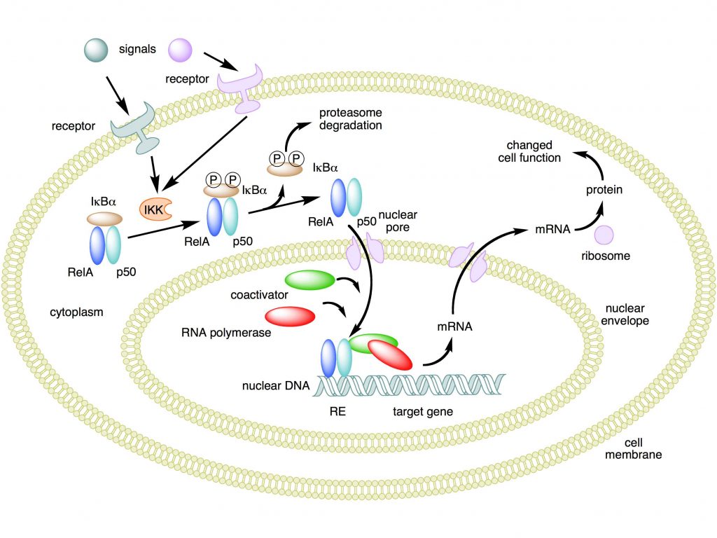
Mechanism of NF-κB action. In this figure, the NF-κB heterodimer consisting of Rel and p50 proteins is used as an example. While in an inactivated state, NF-κB is located in the cytosol complexed with the inhibitory protein IκBα. Through the intermediacy of integral membrane receptors, a variety of extracellular signals can activate the enzyme IκB kinase (IKK). IKK, in turn, phosphorylates the IκBα protein, which results in ubiquitination, dissociation of IκBα from NF-κB, and eventual degradation of IκBα by the proteasome. The activated NF-κB is then translocated into the nucleus where it binds to specific sequences of DNA called response elements (RE). The DNA/NF-κB complex then recruits other proteins such as coactivators and RNA polymerase, which transcribe downstream DNA into mRNA. In turn, mRNA is translated into protein, resulting in a change of cell function.

Attribution: Boghog2 at English Wikipedia [Public domain]

Now, the purpose of this OP is to show, in greater detail, how this mechanism, apparently moderately simple, is indeed extremely complex and dynamic. Let’s see.

The stimuli.

First of all, we must understand what are the stimuli that, arriving to the cell membrane, are capable to activate the NF-kB system. IOWs, what are the signals that work as inputs.

The main concept is: the NF-kB system is a central pathway activated by many stimuli:

  1. Inflammation
  2. Stress
  3. Free
    radicals
  4. Infections
  5. Radiation
  6. Immune
    stimulation

IOWs, a wide variety of aggressive stimuli can activate the system

The extracellular signal arrives to the cell usually through specific cytokines, for example TNF, IL1, or through pathogen associated molecules, like bacterial lipopolysaccharides (LPS). Of course there are different and specific membrane receptors, in particular IL-1R (for IL1) , TNF-R (for TNF), and many TLRs (Toll like receptors, for pathogen associated structures). A special kind of activation is implemented, in B and T lymphocytes, by the immune activation of the specific receptors for antigen epitopes (B cell receptor, BCR, and T cell receptor, TCR).

The process through which the activated receptor can activate the NF-kB dimer is rather complex: it involves, in the canonical pathway, a macromolecular complex called IKK (IkB kinase) complex, comprising two catalytic kinase subunits (IKKa and IKKb) and a regulatory protein (IKKg/NEMO), and involving in multiple and complex ways the ubiquitin system. The non canonical pathway is a variation of that. Finally, a specific protein complex (CBM complex or CBM signalosome) mediates the transmission from the immune BCR or TCR to the canonical pathway. See Fig. 2:

From: NF-κB Activation in Lymphoid Malignancies: Genetics, Signaling, and Targeted Therapy – Scientific Figure on ResearchGate. Available from: https://www.researchgate.net/figure/Increased-activity-of-the-CARMA1-BCL10-MALT1-signalosome-drives-constitutive-NF-kB_fig2_324089636 [accessed 10 Jul, 2019]
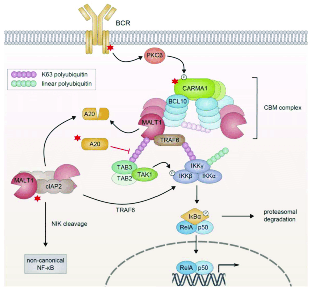
Figure 3 – NF-κB Activation in Lymphoid Malignancies: Genetics, Signaling, and Targeted Therapy
available via license: Creative Commons Attribution 4.0 International

I will not go into further details about this part, but those interested can have a look at this very good paper:

TLR-4, IL-1R and TNF-R signaling to NF-kB: variations on a common theme

In particular, Figg. 1, 2, 3.

In the end, as a result of the activation process, the IkB inhibitor is degraded by the ubiquitin system, and the NK-kB dimer is free to migrate to the nucleus.

An important concept is that this is a “rapid-acting” response system, because the dimers are already present, in inactive form, in the cytoplasm, and must not be synthesized de novo: so the system is ready to respond to the activating signal.

The response.

But what is the cellular response?

Again, there are multiple and complex possible responses.

Essentially, this system is a major regulator of innate and adaptive immune responses. As such, it has a central role in the regulation of inflammation, in immunity, in autoimmune processes, and in cancer.

Moreover, the NF-kB system is rather ubiquitous, and is present and active in many different cell types. And, as we have seen, it can be activated by different stimuli, in different ways.

So, the important point is that the response to activation must be (at least):

  1. Lineage-specific
  2. Stimulus-specific

IOWs, different cells must be able to respond differently, and each cell type must respond differently to different stimuli. That gives a wide range of possible gene expression patterns at the transcription level.

The following paper is a good review of the topic:

Selectivity of the NF-κB Response

For example, IL2 is induced by NF-kB activayion in T cells, but not in B cells (lineage specific response). Moreover, specific cell types can undergo specific, and often different, cell destinies after NF-kB activation: for example, NK-kB is strongly involved in the control and regulation of T and B cell development.

From:

30 years of NF-κB: a blossoming of relevance to human pathobiology

“B and T lymphocytes induce NF-κB in adaptive immune responses through the CARD11:Bcl10:MALT1 (CBM) complex (Hayden and Ghosh, 2008). Newly expressed genes promote lymphocyte proliferation and specific immune functions including antibody production by B cells and the generation of cytokines and other anti-pathogen responses by T cells.”

And, in the same cell type, certain promoters regulated by NF-kB require additional signaling (for example,  in human dendritic cells promoters for Il6Il12b, and MCP-1 require additional p38 histone phosphorylation to be activated), while others can be activated directly (stimulus-specific response).

So, to sum up:

  1. A variety of stimuli can activate the system in different ways
  2. The system itself has its complexity (different dimers)
  3. The response can be widely different, according to the cell type where it happens, and to the type of stimuli that have activated the system, and probably according to other complex variables.
  4. The possible responses include a wide range of regulations of inflammation, of the immune system, of cell specifications or modifications, and so on.

How does it work?

So, what do we know about the working of such a system?

I will ignore, for the moment, the many complexities of the activation pathways, both canonical and non canonical, the role of cyotkines and receptors and IKK complexes, the many facets of NEMO and of the involvement of the ubiquitin system.

For simplicity, we will start with the activated system: the IkB inhibitor has been released from the inactive complex in the cytoplasm, and some form of NF-kB dimer is ready to migrate to the nucleus.

Let’s remember that the purpose of this OP is to show that the system works as a dynamic, far from equilibrium system, rather than as a “traditional” machine. And that such a way to work is an even more amazing example of design and functional complexity.

To do that; I will rely mainly on the recent paper quoted at the beginning:

Considering Abundance, Affinity, and Binding Site Availability in the NF-kB Target Selection Puzzle

The paper is essentially about the NF-kB Target Selection Puzzle. IOWs, it tries to analyze what we know about the specificity of the response. How are specific patterns of transcription achieved after the activation of the system? What mechanisms allow the selection of the right genes to be transcribed (the targets) to implement the specific patterns according to cell type, context, and type of stimuli?

A “traditional” view of the system as a machine would try to establish rather fixed connections. For example, some type of dimer is connected to specific stimuli, and evokes specific gene patterns. Or some other components modulate the effect of NK-kB, generate diversification and specificity of the response.

Well, those ideas are not completely wrong. In a sense, the system does work also that way. Dimer specificity has a role. Other components have a role. In a sense, but only in a sense, the system works as though it were a traditional machine, and uses some of the mechanisms that we find in the concept of a traditional biological machine.

But that is only a tiny part of the real thing.

The real thing is that the system really works as a dynamic, far from equilibrium system, harnessing huge random/stochastic components to achieve robustness and complexity and flexibility of behavior in spite of all those non finalistic parts.

Let’s see how that happens, at least for the limited understanding we have of it. It is important to consider that this is a system that has been studied a lot, for decades, because of its central role in so many physiological and pathological contexts, and so we know many things. But still, our understanding is very limited, as you will see.

So, let’s go back to the paper. I will try to summarize as simply as possible the main concepts. Anyone who is really interested can refer to the paper itself.

Essentially, the paper analyzes three important and different aspects that contribute to the selection of targets at the genomic level by our TFs (IOWs, our NF-kB dimers, ready to migrate to the nucleus. As the title itself summarizes, they are:

  1. Abundance
  2. Affinity
  3. Binding site availability

1. Abundance

Abundance is referred here to two different variables: abundance of NF-kB Binding Sites in the genome and abundance of Nucleus-Localized NF-kB Dimers. Let’s consider them separately.

1a) Abundance of NF-kB Binding Sites in the genome:

It is well known that TFs bind specific sites in the genome. For NF-kB TFs, the following consensus kB site pattern has been found:

 5′-GGGRNWYYCC-3′

where R, W, Y, and N, respectively denote purine, adenine or thymine, pyrimidine, and any nucleotide.

That simply means that any sequence corresponding to that pattern in the genome can, in principle, bind NF-kB dimers.

So the problem is: how many such sequences do exist in the human genome?

Well, a study based on RelA has evaluated about 10^4 consensus sequences in the whole genome, but as NF-kB dimers seem to bind even incomplete consensus sites, the total number of potential binding sites could be nearer to 10^6

1b) Abundance of Nucleus-Localized NF-kB Dimers:

An estimate of the abundance of dimers in the nucleus after activation of the system is that about 1.5 × 10^5 molecules can be found, but again that is derived from studies about RelA only. Moreover, the number of molecules and type of dimer can probably vary much according to cell type.

So, the crucial variable, that is the ratio between binding sites and available dimers, and which could help undertsand the rate of sites saturation in the nucleus, remains rather undecided, and it seems very likely that it can vary a lot in different circumstances.

But there is another very interesting aspect about the concentration of dimers in the nucleus. According to some studies, NF-kB seems to generate oscillations of its nuclear content in some cell types, and those oscillation can be a way to generate specific transcription patterns:

NF-kB oscillations translate into functionally related patterns of gene expression

For example, this very recent paper :

NF-κB Signaling in Macrophages: Dynamics, Crosstalk, and Signal Integration

shows at Fig. 3 the occupancy curve of binding sites at nuclear level after NF-kB activation in two different cell types.

In fibroblasts, the curve is a periodic oscillation, with a frequency that varies according to various factors, and translates into different transcription scenarios accordingly:

Gene expression dynamics scale with the period (g1) and amplitude (g2) of these oscillations, which are influenced by variables such as signal strength, duration, and receptor identity.


In macrophages, instead, the curve is rather:

a single, strong nuclear translocation event which persists for as long as the stimulus remains and tends to remain above baseline for an extended period of time.

In this case, the type of transcription will be probably regulated by the are under the curve, ratehr than by the period and amplitude of the oscialltions, as happened in fibroblasts.

Interestingly, while in previous studies it seemed that the concentration of nuclear dimers could be sufficient to saturate most or all binding sites, that has been found not to be the case in more recent studies. Again from the paper about abundance:

in fact, this lack of saturation of the system is necessary to generate stimulus- and cell-type specific gene expression profiles

Moreover, the binding itself seems to be rather short-lived:

Interestingly, it is now thought that most functional NF-kB interactions with chromatin—interactions that lead to a change in transcription—are fleeting… a subsequent study using FRAP in live cells expressing RelA-GFP showed that most RelA-DNA interactions are actually quite dynamic, with half-lives of a few seconds… Indeed, a recent study used single-molecule tracking of individual Halo-tagged RelA molecules in live cells to show that the majority (∼96%) of RelA undergoes short-lived interactions lasting on average ∼0.5 s, while just ∼4% of RelA molecules form more stable complexes with a lifetime of ∼4 s.

2. Affinity

Affinity of dimers for DNA sequences is not a clear cut matter. From the paper:

Biochemical DNA binding studies of a wide variety of 9–12 base-pair sequences have revealed that different NF-kB dimers bind far more sequences than previously thought, with different dimer species exhibiting specific but overlapping affinities for consensus and non-consensus kB site sequences.

IOWs, we have different dimers (15 different types) binding with varying affinity different DNA sequences (starting from the classical consensus sequence, but including also incomplete sequences). Remember that those sequences are rather short (the consensus sequence is 10 nucleotides long), and that there are thousands of such sequences in the genome.

Moreover, different bindings can affect transcription differently. Again, from the paper:

How might different consensus kB sites modulate the activity of the NF-kB dimers? Structure-function studies have shown that binding to different consensus kB sites can alter the conformation of the bound NF-kB dimers, thus dictating dimer function When an NF-kB dimer interacts with a DNA sequence, side chains of the amino  acids located in the DNA-binding domains of dimers contact the bases exposed in the groove of the DNA. For different consensus kB site sequences different bases are exposed in this groove, and NF-kB seems to alter its conformation to maximize interactions with the DNA and maintain high binding affinity. Changes in conformation may in turn impact NF-kB binding to co-regulators of transcription, whether these are activating or inhibitory, to specify the strength and dynamics of the transcriptional response. These findings again highlight how the huge array of kB binding site sequences must play a key role in modulating the transcription of target genes.

Quite a complex scenario, I would say!

But there is more:

Finally, as an additional layer of dimer and sequence-specific regulation, each of the subunits can be phosphorylated at multiple sites with, depending on the site, effects on nearly every step of NF-kB activation.

IOWs, the 15 dimers we have mentioned can be phosphorylated in many different ways, and that changes their binding affinities and their effects on transcription.

This section of the paper ends with a very interesting statement:

Overall, when considering the various ways in which NF-kB dimer abundances and their affinity for DNA can be modulated, it becomes clear that with these multiple cascading effects, small differences in consensus kB site sequences and small a priori differences in interaction affinities can ultimately have a large impact on the transcriptional response to NF-kB pathway activation.

Emphasis mine.

This is interesting, because in some way it seems to suggest that the whole system acts like a chaotic system, at least at some basic level. IOWs, small initial differences, maybe even random noise, can potentially affect deeply the general working of the whole systems.

Unless, of course, there is some higher, powerful level of control.

3. Availability of high affinity kB binding sequences

We have seen that there is a great abundance and variety of binding sequences for NF-kB dimers in the human genome. But, of course, those sequences are not necessarily available. Different cell types will have a different scenario of binding sites availability.

Why?

Because, as we know, the genome and chromatin are a very dynamic system, that can exist in many different states, continuosly changing in different cell types and, in the same cell type, in different conditions..

We know rather well the many levels of control that affect DNA and chromatin state. In brief, they are essentially:

  1. DNA methylation
  2. Histone modifications (methylation, acetylation, etc)
  3. Chromatin modifications
  4. Higher levels of organization, including nuclear localization and TADs (Topologically Associating Domains)

For example, from the paper:

The promoter regions of early response genes have abundant histone acetylation or trimethylation prior to stimulation [e.g., H3K27ac, (67) and H4K20me3, (66)], a chromatin state “poised” for immediate activation…  In contrast, promoters of late genes often have hypo-acetylated histones, requiring conformational changes to the chromatin to become accessible. They are therefore unable to recruit NF-kB for up to several hours after stimulation (68), due to the slow process of chromatin remodeling.

We must remember that each wave of NK-kB activation translates into the modified transcription of a lot of different genes at the genome level. It is therefore extremely important to consider what genes are available (IOWs, their promoters can be reached by the NF-kB signal) in each cell type and cell state.

The paper concludes:

Taken together, chromatin state and chromatin organization strongly influence the selection of DNA binding sites by NF-kB dimers and, most likely, the selection of the target genes that are regulated by these protein-DNA interaction events. Analyses that consider binding events in the context of three-dimensional nuclear organization and chromatin composition will be required to generate a more accurate view of the ways in which NF-kBDNA binding affects gene transcription.

This is the main scenario. But there are other components, that I have not considered in detail for the sake of brevity, for example competition between NF-kB dimers and the complex role and intervention of other co-regulators of transcription.

Does the system work?

But does the system work?

Of course it does. It is a central regulator, as we have said, of many extremely important biological processes, above all immunity. This is the system that decides how immune cells, T and B lymphocytes, have to behave, in terms of cell destiny and cell state. It is of huge relevance in all inflammatory responses, and in our defense against infections. It works, it works very well.

And what happens if it does not work properly?

Of course, like all very complex systems, errors can happen. Those interested can have a look at this recent paper:

30 years of NF-κB: a blossoming of relevance to human pathobiology

First of all, many serious genetic diseases have been linked to mutations in genes involved in the system. You can find a list in Table 1 of the above paper. Among them, for example, some forms of SCID, Severe combined immunodeficiency, one of the most severe genetic diseases of the immune system.

But, of course, a dysfunction of the NF-kB system has a very important role also in autoimmune diseases and in cancer.

Conclusions.

So, let’s try to sum up what we have seen here in the light of the original statement about biological systems that “are not machines”.

The NF-kB system is a perfect example. Even if we still understand very little of how it works, it is rather obvious that it is not a traditional machine.

A traditional machine would work differently. The signal would be transmitted from the membrane to the nucleus in the simplest possible way, without ambiguities and diversions. The Transcription Factor, once activated, would bind, at the level of the genome, very specific sites, each of them corresponding to a definite cascade of specific genes. The result would be clear cut, almost mechanical. Like a watch.

But that’s not the way things happen. There are myriads of variations, of ambiguities, of stochastic components.

The signal arrives to the membrane in multiple ways, very different one from the other: IL1, IL17, TNF, bacterial LPS, and immune activation of the B cell receptor (BCR) or the T cell receptor (TCR) are all possible signals.

The signal is translated to the NF-kB proteins in very different ways: canonical or non canonical activation, involving complex protein structures such as:

The CBM signalosome, intermediate between immune activation of BCR or TCR and canonical activation of the NF-kB. This complex is made of at least three proteins, CARD11, Bcl10 and MALT1.

The IKK complex in canonical activation: this is made of three proteins, IKK alpha, IKK beta, and NEMO. Its purpose is to phosphorylate the IkB, the inhibitor of the dimers, so that it can be ubiquinated and released from the dimer. Then the dimer can relocate to the nucleus.

Non canonical pathway: it involves the following phosphorylation cascade: NIK -> IKK alpha dimer -> Relb – p100 dimer -> Relb – p50 dimer (the final TF). It operates during the development of lymphoid organs and is responsible for the generation of B and T lymphocytes.

Different kinds of activated dimers relocate to the nucleus.

Different dimers, in varying abundance, interact with many different binding sites: complete or incomplete consensus sites, and probably others. The interaction is usually brief, and it can generate an oscillating pattern, or a more stable pattern

Completely different sets of genes are transcribed in different cell types and in different contexts, because of the interaction of NF-kB TFs with their promoters.

Many other factors and systems contribute to the final result.

The chromatin state of the cell at the moment of the NF-kB activation is essential to determine the accessibility of different binding sites, and therefore the final transcription pattern.

All these events and interactions are quick, unstable, far from equilibrium. A lot of possible random noise is involved.

In spite of that amazing complexity and potential stochastic nature of the system, reliable transcripion regulation and results are obtained in most cases. Those results are essential to immune cell differentiation, immune response, both innate and adaptive, inflammation, apoptosis, and many other crucial cellular processes.

So, let’s go back to our initial question.

Is this the working of a machine?

Of course it is! Because the results are purposeful, reasonably robust and reliable, and govern a lot of complex processes with remarkable elegance and efficiency.

But certainly, it is not a traditional machine. It is a lot more complex. It is a lot more beautiful and flexible.

It works with biological realities and not with transistors and switches. And biological realities are, by definition, far from equilibrium states, improbable forms of order that must continuously recreate themselves, fighting against the thermodynamic disorder and the intrinsic random noise that should apparently dominate any such scenario.

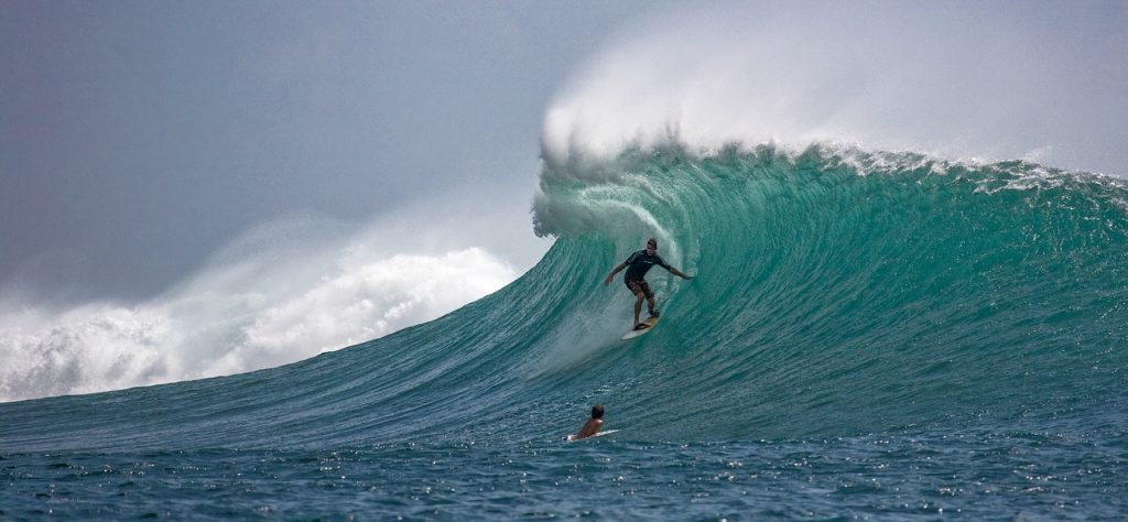
It is more similar to a set of extremely clever surfers who succeed in performing elegant and functional figures and motions in spite of the huge contrasting waves.

It is, from all points of view, amazing.

Now, Paley was absolutely right. No traditional machine, like a watch, could ever originate without design.

And if that is true of a watch, with its rather simple and fixed mechanisms, how much truer it must be for a system like NF-kB? Or, for that, like any cellular complex system?

Do you still have any doubts?

Added graphic: The evolutionary history, in terms of human conserved information, of the three proteins in the CBM signalosome.
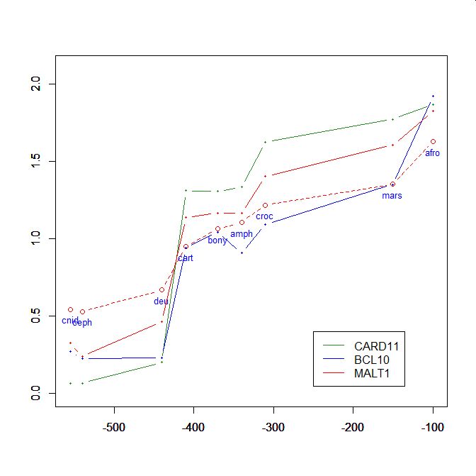
On the y axis, homologies with the human protein as bits per aminoacid (bpa). On the x axis, approximate time of appearance in million of years.
The graphic shows the big information jump in vertebrates for all three protens , especially CARD11.


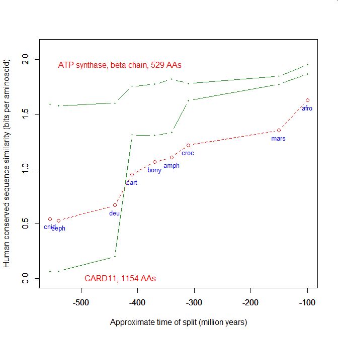
Added graphic: two very different proteins and their functional history


Added graphic (for Bill Cole). Functional history of Prp8, collagen, p53.
Comments
Sven Mil at #267: You really don't understand, do you? My method (blast alignment by the default algorithm) is simply the method used routinely by almost all researchers to detect sequence homology. So, I am not doing anything particular, as you seem to believe. Those who are interested in detecting weak and distant homologies, of course, can use other methods, like more sensitive algorithms of alignment and structural similarities, if they like. That will give higher sensitivity and lower specificity in detecting if two proteins are homologues. IOWs, more false positives. As I have said many times, I am not trying to detect if two proteins are distant homologues, because that has nothing to do with my reasoning. The researchers you quote say that sigma factor and human TFIIB are homologues? Maybe. Maybe not. Anyway, I have no problems with that statement. If they are, they are. That makes no difference in my reasoning. More in next post.gpuccio
August 4, 2019
August
08
Aug
4
04
2019
12:21 AM
12
12
21
AM
PDT
GP, Sven Mil, ET, et al, I’m ignorant of basic biology. I’ve tried to understand what you’re discussing but can’t figure it out. Please, explain this to me in easy to understand terms: 1. Are you comparing two proteins P1 and P2 which work for prokaryotes (P1) and eukaryotes (P2) respectively? 2. Could P1 work for eukaryotes too? 2.1. If YES then why wasn’t it kept in eukaryotes rather than being replaced by P2? 3. Any idea how P1 and P2 could have appeared? I may have more questions, but these are fine to start. Note that I would like to read the answers from all of you and from other readers of this discussion. Thanks.PeterA
August 3, 2019
August
08
Aug
3
03
2019
08:31 PM
8
08
31
PM
PDT
sven mil:
The fact is Gpucc, you are unable to detect homology between two proteins that perform the same function and that have been shown to be homologs by other methods.
How are using the word "homology"? Convergence explains two different proteins having the same function, as does a common design.
You can detect high conservation (i.e. when a protein’s functional niche has become well-defined and locked into place evolutionarily speaking) but you are completely unable to detect the actual evolution of a protein
Is there any evidence that blind and mindless processes can produce proteins? I would think that gpuccio is open to the concept of proteins evolving by means of intelligent design.ET
August 3, 2019
August
08
Aug
3
03
2019
03:04 PM
3
03
04
PM
PDT
The fact is Gpucc, you are unable to detect homology between two proteins that perform the same function and that have been shown to be homologs by other methods. This means your method is simply not sensitive enough (as you have already admitted) to trace the evolution of proteins back in the way that you are attempting to. You can detect high conservation (i.e. when a protein's functional niche has become well-defined and locked into place evolutionarily speaking) but you are completely unable to detect the actual evolution of a protein And that's why you will always find your "jumps" if you go back far enough. You seem smart enough that I'd bet you knew that from the start... Guess I shouldn't really be surprisedSven Mil
August 3, 2019
August
08
Aug
3
03
2019
02:32 PM
2
02
32
PM
PDT
Widespread roles of enhancer-like transposable elements in cell identity and long-range genomic interactions https://www.ncbi.nlm.nih.gov/pmc/articles/PMC6314169/OLV
August 3, 2019
August
08
Aug
3
03
2019
12:49 PM
12
12
49
PM
PDT
GP, The increasing number of research papers on this OP topic definitely point to complex functional information processing systems with multiple control levels that can only result from conscious design. Please, I would like to read your comments on any of the papers linked @264 that you haven’t cited before. Thanks.OLV
August 3, 2019
August
08
Aug
3
03
2019
03:42 AM
3
03
42
AM
PDT
GP, There’s so much literature on transcription regulation that it’s difficult to look at them all. Here’s just a small sample: (Note that you have cited some of these papers) Transcription-driven chromatin repression of Intragenic transcription start sites https://www.ncbi.nlm.nih.gov/pmc/articles/PMC6373976/ Genome-wide enhancer annotations differ significantly in genomic distribution, evolution, and function https://www.ncbi.nlm.nih.gov/pmc/articles/PMC6585034/ Computational Biology Solutions to Identify Enhancers-target Gene Pairs https://www.ncbi.nlm.nih.gov/pmc/articles/PMC6611831/ Detection of condition-specific marker genes from RNA-seq data with MGFR https://www.ncbi.nlm.nih.gov/pmc/articles/PMC6542349/ Enhancer RNAs: Insights Into Their Biological Role https://www.ncbi.nlm.nih.gov/pmc/articles/PMC6505235/ Epigenetic control of early dendritic cell lineage specification by the transcription factor IRF8 in mice http://www.bloodjournal.org/content/133/17/1803.long?sso-checked=true Competitive endogenous RNA is an intrinsic component of EMT regulatory circuits and modulates EMT https://www.ncbi.nlm.nih.gov/pmc/articles/PMC6456586/ Delta Like-1 Gene Mutation: A Novel Cause of Congenital Vertebral Malformation https://www.ncbi.nlm.nih.gov/pmc/articles/PMC6593294/OLV
August 3, 2019
August
08
Aug
3
03
2019
03:11 AM
3
03
11
AM
PDT
jawa, That discussion is over. GP took care of it appropriately and wisely continued to provide very interesting information on the current topic. This thread has already exceeded my expectations.PeterA
July 31, 2019
July
07
Jul
31
31
2019
03:15 AM
3
03
15
AM
PDT
OLV @261: Maybe Sven Mil can help to answer your questions, after he responds the GP’s comments addressed to him after his last comment @240? :)jawa
July 31, 2019
July
07
Jul
31
31
2019
02:55 AM
2
02
55
AM
PDT
GP @256: “It seems perfectly natural that a polymorphic semiotic system like NF-kB is strictly regulated by another universal semiotic system, the ubiquitin system. And the regulation is not simple at all, but deeply and semiotically complex:” Is it also natural that those semiotic systems resulted from natural selection operating on random variations over gazillion years? I’m looking for the literature where this is explained. For example, what did those systems evolve from? What were their ancestors?OLV
July 31, 2019
July
07
Jul
31
31
2019
02:42 AM
2
02
42
AM
PDT
GP @257: “orchestrate and fine-tune cellular metabolism at various levels of operation.” “A new paradigm in fine tuning? We are becoming accustomed to that kind of thing,” Agree.OLV
July 31, 2019
July
07
Jul
31
31
2019
02:07 AM
2
02
07
AM
PDT
GPuccio, Definitely you’re on a roll! You’ve referenced several very interesting papers in a row.OLV
July 30, 2019
July
07
Jul
30
30
2019
08:17 PM
8
08
17
PM
PDT
To all: Another rather exotic level of regulation of the NF-kB system: immunophilins. Regulation of NF-kB signalling cascade by immunophilins http://www.eurekaselect.com/131456/article
Abstract: The fine regulation of signalling cascades is a key event required to maintain the appropriate functional properties of a cell when a given stimulus triggers specific biological responses. In this sense, cumulative experimental evidence during the last years has shown that high molecular weight immunophilins possess a fundamental importance in the regulation of many of these processes. It was first discovered that TPR-domain immunophilins such as FKBP51 and FKBP52 play a cardinal role, usually in an antagonistic fashion, in the regulation of several members of the steroid receptor family via its interaction with the heat-shock protein of 90-kDa, Hsp90. These Hsp90-associated cochaperones form a functional unit with the molecular chaperone influencing ligand binding capacity, receptor trafficking, and hormone-dependent transcriptional activity. Recently, it was demonstrated that the same immunophilins are also able to regulate the NF-kB signalling cascade in an Hsp90 independent manner. In this article we analize these properties and discuss the relevance of this novel regulatory pathway in the context of the pleiotropic actions managed by NF-kB in several cell types and tissues.
Emphasis mine. You may rightfully ask: what are immunophilins? Let's take a simple answer from Wikipedia: "immunophilins are endogenous cytosolic peptidyl-prolyl isomerases (PPI) that catalyze the interconversion between the cis and trans isomers of peptide bonds containing the amino acid proline (Pro). They are chaperon molecules that generally assist in the proper folding of diverse "client" proteins". Here is a recent review about them: Biological Actions of the Hsp90-binding Immunophilins FKBP51 and FKBP52 https://www.ncbi.nlm.nih.gov/pmc/articles/PMC6406450/
Abstract: Immunophilins are a family of proteins whose signature domain is the peptidylprolyl-isomerase domain. High molecular weight immunophilins are characterized by the additional presence of tetratricopeptide-repeats (TPR) through which they bind to the 90-kDa heat-shock protein (Hsp90), and via this chaperone, immunophilins contribute to the regulation of the biological functions of several client-proteins. Among these Hsp90-binding immunophilins, there are two highly homologous members named FKBP51 and FKBP52 (FK506-binding protein of 51-kDa and 52-kDa, respectively) that were first characterized as components of the Hsp90-based heterocomplex associated to steroid receptors. Afterwards, they emerged as likely contributors to a variety of other hormone-dependent diseases, stress-related pathologies, psychiatric disorders, cancer, and other syndromes characterized by misfolded proteins. The differential biological actions of these immunophilins have been assigned to the structurally similar, but functionally divergent enzymatic domain. Nonetheless, they also require the complementary input of the TPR domain, most likely due to their dependence with the association to Hsp90 as a functional unit. FKBP51 and FKBP52 regulate a variety of biological processes such as steroid receptor action, transcriptional activity, protein conformation, protein trafficking, cell differentiation, apoptosis, cancer progression, telomerase activity, cytoskeleton architecture, etc. In this article we discuss the biology of these events and some mechanistic aspects.
In particular, section 6: "Immunophilins Regulate NF-kB Activity"gpuccio
July 30, 2019
July
07
Jul
30
30
2019
08:42 AM
8
08
42
AM
PDT
To all: Oh, this is really new. Did you know that TFs seem to have a key role not only in nuclear transcription regulation, but also in the regulation of those other strange genome-bearing organelles, the mitochondria? Of course, NF-kB is one of the TF systems involved there, too: Nuclear Transcription Factors in the Mitochondria: A New Paradigm in Fine-Tuning Mitochondrial Metabolism. https://www.ncbi.nlm.nih.gov/pubmed/27417432
Abstract: Noncanonical functions of several nuclear transcription factors in the mitochondria have been gaining exceptional traction over the years. These transcription factors include nuclear hormone receptors like estrogen, glucocorticoid, and thyroid hormone receptors: p53, IRF3, STAT3, STAT5, CREB, NF-kB, and MEF-2D. Mitochondria-localized nuclear transcription factors regulate mitochondrial processes like apoptosis, respiration and mitochondrial transcription albeit being nuclear in origin and having nuclear functions. Hence, the cell permits these multi-stationed transcription factors to orchestrate and fine-tune cellular metabolism at various levels of operation. Despite their ubiquitous distribution in different subcompartments of mitochondria, their targeting mechanism is poorly understood. Here, we review the current status of mitochondria-localized transcription factors and discuss the possible targeting mechanism besides the functional interplay between these factors.
Emphasis mine. A new paradigm in fine tuning? We are becoming accustomed to that kind of thing, I suppose! :)gpuccio
July 30, 2019
July
07
Jul
30
30
2019
01:26 AM
1
01
26
AM
PDT
To all: It seems perfectly natural that a polymorphic semiotic system like NF-kB is strictly regulated by another universal semiotic system, the ubiquitin system. And the regulation is not simple at all, but deeply and semiotically complex: The Met1-Linked Ubiquitin Machinery: Emerging Themes of (De)regulation https://www.cell.com/molecular-cell/fulltext/S1097-2765(17)30649-4?_returnURL=https%3A%2F%2Flinkinghub.elsevier.com%2Fretrieve%2Fpii%2FS1097276517306494%3Fshowall%3Dtrue
The linear ubiquitin chain assembly complex, LUBAC, is the only known mammalian ubiquitin ligase that makes methionine 1 (Met1)-linked polyubiquitin (also referred to as linear ubiquitin). A decade after LUBAC was discovered as a cellular activity of unknown function, there are now many lines of evidence connecting Met1-linked polyubiquitin to NF-?B signaling, cell death, inflammation, immunity, and cancer. We now know that Met1-linked polyubiquitin has potent signaling functions and that its deregulation is connected to disease. Indeed, mutations and deficiencies in several factors involved in conjugation and deconjugation of Met1-linked polyubiquitin have been implicated in immune-related disorders. Here, we discuss current knowledge and recent insights into the role and regulation of Met1-linked polyubiquitin, with an emphasis on the mechanisms controlling the function of LUBAC. Main Text: Transcription factors in the nuclear factor-kB (NF-kB) family orchestrate inflammatory responses and their activation by immune receptors, such as pattern recognition receptors (PRRs), cytokine receptors, and antigen receptors, is important for innate and adaptive immune function. A unifying feature of the signaling processes triggered by these receptors is that they rely on formation of ubiquitin (Ub) chains to transmit the signal from the activated receptor to the nucleus for stimulation of NF-kB-mediated transcription (Figure 1). The discovery that Ub chains are required for NF-kB activation was reported more than 20 years ago with the finding that Inhibitor of NF-kB alpha (IkBalpha, also termed NFKBIA) is modified with Ub chains (linked via Lys48; Lys48-Ub) in response to receptor activation, leading to its rapid degradation via the proteasome (Chen et al., 1995, Palombella et al., 1994, Traenckner et al., 1994). Subsequently, a series of studies by Zhijian “James” Chen and colleagues showed that Ub chains linked via Lys63 (Lys63-Ub) play a non-degradative role in kinase signaling and NF-kB activation by facilitating the activation of transforming growth factor (TGF)-beta-activated kinase 1 (TAK1) (Deng et al., 2000, Wang et al., 2001). In 2006, Kazuhiro Iwai and colleagues identified a Ub E3 ligase complex that only assembles Ub chains through the N-terminal methionine (Met1-Ub); they called this the linear Ub chain assembly complex (LUBAC) and subsequently discovered that LUBAC stimulates NF-kB activity by conjugating Met1-Ub (Tokunaga et al., 2009, Kirisako et al., 2006). Now, after 10 years of research into LUBAC and Met1-Ub biology, it is clear that Met1-Ub harbors potent signaling properties and, together with Lys63-Ub and Lys48-Ub, plays a central role in NF-kB activation and immune function (Figure 2). Met1-Ub is also implicated in signaling by viral nucleotide-sensing receptors, leading to interferon response factor (IRF)-mediated transcription (Figure 1) and other signaling processes (reviewed in Sasaki and Iwai, 2015). In this review, we primarily discuss its role in NF-kB signaling.
gpuccio
July 30, 2019
July
07
Jul
30
30
2019
01:10 AM
1
01
10
AM
PDT
This discussion is the third most visited the last 30 days! Definitely a fascinating topic. Congratulations to GP!jawa
July 29, 2019
July
07
Jul
29
29
2019
08:22 PM
8
08
22
PM
PDT
GPuccio @253: Excellent explanation. Thanks!OLV
July 29, 2019
July
07
Jul
29
29
2019
08:14 PM
8
08
14
PM
PDT
Sven Mil, OLV and all: Some more facts: 1) The archaeal TFB shows definite and highly significant sequence homology with human TFIIB. These are the results of the usual Blast alignment, always using the defaul algorithm and nothing else: Proteins: Human general TFIIB (Q00403) vs archaeal TFB (A0A2D6Q6B7): Identities: 93; Positives: 154; Bitscore: 172 bits; E value: 2e-56 2) No significant sequence homology can be detected instead, using the same identical methodology, between bacterial sigma factor 70 and the archeal TFB: Proteins: Sigma factor 70 E. coli (P00579) vs archaeal TFB (A0A2D6Q6B7): Identities: 25; Positives: 44; Bitscore: 16.2 bits; E value: 2.0 3) And, of course, as already said, no significant sequence homology can be detected, using the same identical methodology, between bacterial sigma factor 70 and human TFIIB: Proteins: Sigma factor 70 E. coli (P00579) vs Human general TFIIB (Q00403): Identities: 32; Positives: 49; Bitscore: 16.9 bits; E value: 1.4 (plus three more non significant short alignments, with E values of 2.5, 2.7, 3.6) These are simple facts, that can be verified by all. At sequence level, there is a definite homology (anyway only partial) between the archaeal protein and the human protein. That corresponds to the well known concept that transcitpion initiation in archaea is much more similar to transcription initiation in eukaryotes, while in bacteria it is very different. Indeed, no significant sequence homology can be detected, always using that same methodology, between the human and the bacterial protein, or between the bacterial and the archaeal protein. These simple facts are undeniable. Check what I have written in my comment #202, to John_a_designer: "Now, in eukaryotes there are six general TFs. Archea have 3. In bacteria sigma factors have the role of general TFs. Sigma factors, archaeal general TFs and eukaryotic general TFs seem to share some homology. I think that the archaeal system, however, is much more similar to the eukaryotic system, and that includes RNA polymerases. ... While archaea are more similar to eukaryotes in the system of general TF, the regulation of transcription by one or two suppressor or activator seems to be similar to what described for bacteria. Finally, there is another important aspect where archaea are more similar to eukarya. Their chromatin structure is based on histones and nucleosome, like in eukaryotes, but the system is rather different from the corresponding eukaryotic system. Instead, bacteria have their form of DNA compression, but it is not based on histones and nucleosomes."gpuccio
July 29, 2019
July
07
Jul
29
29
2019
01:42 PM
1
01
42
PM
PDT
GPuccio, It’s my pleasure to post links to interesting papers that sometimes I find in different journals. In some cases they may shed more light on the discussed topics.OLV
July 29, 2019
July
07
Jul
29
29
2019
12:59 PM
12
12
59
PM
PDT
OLV: Thank you for the very interesting links. Yes, we are certainly not even near to a real understanding of how transcription is regulated. More on these fascinating topics as soon as I can use again a true keyboard! :)gpuccio
July 29, 2019
July
07
Jul
29
29
2019
07:54 AM
7
07
54
AM
PDT
Biology research seems like a never-ending story: The more we know, more is there for us to learn from. Really fascinating, isn’t it?
Shaking the dogma “These results question the generality of a current dogma in the field, that chromatin domains (TADs) are essential to constrain and restrict enhancer function,” says Eileen Furlong, the EMBL group leader who led the study. “We were able to show that major changes in the 3D organisation of the genome had surprisingly little effect on the expression of most genes, at least in this biological context. The results indicate that while some genes are affected, many appear resistant to rearrangements in their chromatin domain, and that only a small fraction of genes are sensitive to such changes in their topology.” Enhancers are not that promiscuous This raises many interesting questions in the field of chromatin topology, for example: what are these other mechanisms that control the interactions between enhancers and their target genes? Many enhancers do not appear to be promiscuous: they do not link to just any target gene, but rather have preferred partners. The team will continue to dissect this by using genetics, optogenetics (a technique to control protein activity with laser light) and single-cell approaches. This will allow them to study the impact of many more perturbations to chromatin topology in both cis and trans.
OLV
July 29, 2019
July
07
Jul
29
29
2019
05:43 AM
5
05
43
AM
PDT
GP, The plot thickens... “changes in chromatin domains were not predictive of changes in gene expression. This means that besides domains, there must be other mechanisms in place that control the specificity of interactions between enhancers and their target genes.” More control mechanisms? Don’t we have enough control mechanisms to keep track of already? :)OLV
July 29, 2019
July
07
Jul
29
29
2019
05:01 AM
5
05
01
AM
PDT
A genome disconnect
Chromatin loops and domains are major organizational hallmarks of chromosomes. New work suggests, however, that these topological features of the genome are poor global predictors of gene activity, raising questions about their function.
  Highly rearranged chromosomes reveal uncoupling between genome topology and gene expression  
Chromatin topology is intricately linked to gene expression, yet its functional requirement remains unclear. Here, we comprehensively assessed the interplay between genome topology and gene expression using highly rearranged chromosomes (balancers) spanning ~75% of the Drosophila genome. Using transheterozyte (balancer/wild-type) embryos, we measured allele-specific changes in topology and gene expression in cis, while minimizing trans effects. Through genome sequencing, we resolved eight large nested inversions, smaller inversions, duplications and thousands of deletions. These extensive rearrangements caused many changes to chromatin topology, disrupting long-range loops, topologically associating domains (TADs) and promoter interactions, yet these are not predictive of changes in expression. Gene expression is generally not altered around inversion breakpoints, indicating that mis-appropriate enhancer–promoter activation is a rare event. Similarly, shuffling or fusing TADs, changing intra-TAD connections and disrupting long-range inter-TAD loops does not alter expression for the majority of genes. Our results suggest that properties other than chromatin topology ensure productive enhancer–promoter interactions.
The plot thickens:   Does rearranging chromosomes affect their function?  OLV
July 29, 2019
July
07
Jul
29
29
2019
12:46 AM
12
12
46
AM
PDT
Dynamic interplay between enhancer–promoter topology and gene activity  
A long-standing question in gene regulation is how remote enhancers communicate with their target promoters, and specifically how chromatin topology dynamically relates to gene activation. Here, we combine genome editing and multi-color live imaging to simultaneously visualize physical enhancer–promoter interaction and transcription at the single-cell level in Drosophila embryos. By examining transcriptional activation of a reporter by the endogenous even-skippedenhancers, which are located 150?kb away, we identify three distinct topological conformation states and measure their transition kinetics. We show that sustained proximity of the enhancer to its target is required for activation. Transcription in turn affects the three-dimensional topology as it enhances the temporal stability of the proximal conformation and is associated with further spatial compaction. Furthermore, the facilitated long-range activation results in transcriptional competition at the locus, causing corresponding developmental defects. Our approach offers quantitative insight into the spatial and temporal determinants of long-range gene regulation and their implications for cellular fates.
 OLV
July 29, 2019
July
07
Jul
29
29
2019
12:37 AM
12
12
37
AM
PDT
Design Principles Of Mammalian Transcriptional Regulation
Transcriptional regulation occurs via changes to different biochemical steps of transcription, but it remains unclear which steps are subject to change upon biological perturbation. Single cell studies have revealed that transcription occurs in discontinuous bursts, suggesting that features of such bursts like burst fraction (what fraction of time a gene spends transcribing RNA) and burst intensity could be points of transcriptional regulation. Both how such features might be regulated and the prevalence of such modes of regulation are unclear. I first used a synthetic transcription factor to increase enhancer-promoter contact at the ?-globin locus. Increasing promoter- enhancer contact specifically modulated the burst fraction of ?-globin in both immortalized mouse and primary human erythroid cells. This finding raised the question of how generally important the phenomenon of burst fraction regulation might be, compared to other modes of regulation. For example, biochemical studies have suggested that stimuli predominantly affect the rate of RNA polymerase II (Pol II) binding and the rate of Pol II release from promoter-proximal pausing, but the prevalence of these modes of regulation compared to changes in bursting had not been examined. I combined Pol II ChIP-seq and single cell transcriptional measurements to reveal that an independently regulated burst initiation step is required before polymerase binding can occur, and that the change in burst fraction produced by increased enhancer-promoter contact was caused by an increased burst initiation rate. Using a number of global and targeted transcriptional regulatory perturbations, I showed that biological perturbations regulated both burst initiation and polymerase pause release rates, but seemed not to regulate polymerase binding rate. Our results suggest that transcriptional regulation primarily acts by changing the rates of burst initiation and polymerase pause release.
The cells of a eukaryotic organism all share the same genome; however, they differentiate from a single zygote into many different cell types that carry out different functions mediated by the expression of cell-type-specific suites of proteins. A major focus of biological  science has been to understand how cells with the same genome can induce and maintain such divergent functional states. Relatedly, eukaryotic cells must be able to respond quickly to certain stimuli by changing protein expression: canonical examples of such stimuli include heat shock or inflammatory signals. Both cell-type identity and functional responses to signaling are chiefly governed at the level of DNA transcription into RNA, though other processes like protein posttranslational modification and degradation also play important roles.
Many unanswered questions related to the regulation of transcriptional bursting persist.
   OLV
July 29, 2019
July
07
Jul
29
29
2019
12:27 AM
12
12
27
AM
PDT
Displacement of the transcription factor B reader domain during transcription initiation Stefan Dexl, Robert Reichelt, Katharina Kraatz, Sarah Schulz, Dina Grohmann, Michael Bartlett, Michael Thomm Nucleic Acids Research, Volume 46, Issue 19, Pages 10066–10081 DOI: 10.1093/nar/gky699
Transcription initiation by archaeal RNA polymerase (RNAP) and eukaryotic RNAP II requires the general transcription factor (TF) B/ IIB. Structural analyses of eukaryotic transcription initiation complexes locate the B-reader domain of TFIIB in close proximity to the active site of RNAP II. Here, we present the first crosslinking mapping data that describe the dynamic transitions of an archaeal TFB to provide evidence for structural rearrangements within the transcription complex during transition from initiation to early elongation phase of transcription. Using a highly specific UV-inducible crosslinking system based on the unnatural amino acid para-benzoyl-phenylalanine allowed us to analyze contacts of the Pyrococcus furiosus TFB B-reader domain with site-specific radiolabeled DNA templates in preinitiation and initially transcribing complexes. Crosslink reactions at different initiation steps demonstrate interactions of TFB with DNA at registers +6 to +14, and reduced contacts at +15, with structural transitions of the B-reader domain detected at register +10. Our data suggest that the B-reader domain of TFB interacts with nascent RNA at register +6 and +8 and it is displaced from the transcribed-strand during the transition from +9 to +10, followed by the collapse of the transcription bubble and release of TFB from register +15 onwards.
OLV
July 28, 2019
July
07
Jul
28
28
2019
06:15 PM
6
06
15
PM
PDT
Sven Mil: As you have tried to make your non arguments more detailed, you certainly deserve a more detailed answer. As at present I can only answer from my phone, I will be brief for the moment (I am very bad at typing on the phone). Tomorrow I should be able to answer in greater length. Your biggest errors (but not the only ones) are: a) Thinking that I am denying that the two proteins are homologues, or evolutionary related. That is completely false. I have simply blasted the two proteins, and found no detectable homology. That is a simple fact. You can blast them too, and you will have the same result. That means that there is no obvious sequence homology using the default blast algorithm. Again, that is a very simple fact. I have also said that the authors of the paper I linked had used a different method, using structural considerations and different alignment algorithms, because they were interested in detecting a weak relationship to find a possible evolutionary relationship. That's perfectly fine, but I have no interest in affirming or denying a possible evolutionary relationship. If the two proteins are evolutionarily related, that's no problem for me. As you know, I believe in Common Descent by design. b) Thinking that two similar functions are identical. I have already discussed that. Just to add a point, of course all proteins that bind DNA, and that includes all TFs, have a DBD. I don't think that makes their functions identical, virtually or not. c) Thinking that I have problems with the idea that two proteins with highly different sequence can have a similar function. I have no problems with that. But the simple fact remains that in most cases proteins that retain a highly similar, maybe almost identical function through billions of years, like the alpha and beta chains of ATP synthase, show high sequence conservation. Look also at histones and ubiquitin, and thousands and thousands of other examples. Nobody who really believes in the basics of modern biology can deny that sequence conservation through long evolutionary periods is a measure of functional constraint. d) Thinking that I can detect only high sequence homologies. That is completely false. I use the default blast algorithm to have always the same tool in measuring sequence homology. And the default blast algorithms detects very well most sequence homologies, both low and high, and gives a definite measure of the relevance of those homologies in statistical terms, the E value. So, when I say that I could find no detectable homology, I mean a very precise fact: that blasting those two sequences, that I have clearly indicated, with the default blast algorithm, no homology is detected that reaches a significant E value. Again, you can blast the two sequences yourself. This is the method commonly used to detect homology between sequences. e) So, my procedure detects sequence homologies, both weak and strong. I am interested in jumps not because I can only detect jumps, as you foolishly seem to suggest, buy because jumps are clear indicators of design. I find a lot of jumps, some of them really big, and I find a lot of non jumps. As my graphics clearly show. For example, as I have argued in this same thread, TFs usually do not show big jumps, for example at the vertebrate level, for two interesting reasons: 1) Their DBDs are highly conserved and very old, older usually than the vertebrate appearances, usually already well detectable in single celled eukaryotes. 2) Their other domains or sequences are usually poorly conserved during the evolutionary history of metazoa. However, there are strong indications that such a sequence diversification is functional, and not simply a case of neutral variation in non functional sequences. I have made this argument here for RelA, at post #29. Well, that is enough for the moment.gpuccio
July 28, 2019
July
07
Jul
28
28
2019
04:12 PM
4
04
12
PM
PDT
– how could two proteins, vastly different in sequence (according to your BLASTing) carry out the same function?
Different binding partners in the function. The different binding partners can change the rate of transcription. You may be comparing a light switch to a light dimmer and not know it. Gpuccio's method measures protein sequence divergence over time showing resistance to change based on purifying selection. This allows you to demonstrate substitutability and therefor genetic information. You first need to understand his method before trying to make an argument. So far you are talking over him. When you compare a eukaryotic cell to a prokaryotic cell you are using apples and oranges for your comparison and your argument fails.bill cole
July 28, 2019
July
07
Jul
28
28
2019
02:28 PM
2
02
28
PM
PDT
This discussion seems interesting, but flies high above my head. What are the main differences between prokaryotic and eukaryotic cells? I tried to search for it but got gazillion results and don't know where to start from. Here are some abbreviations used in this discussion: BRE TFB/TFIIB recognition element CLR/HTH cyclin-like repeat/helix-turn-helix domain DPBB double psi beta barrel DDRP DNA-dependent RNA polymerase GTF general transcription factor LECA last eukaryotic common ancestor LUCA last universal common ancestor Ms Methanocaldococcus sp. FS406-22, PIF primordial initiation factor RDRP RNA-dependent RNA polymerase RNAP RNA polymerase Sc Saccharomyces cerevisiae TFB transcription factor B TFIIB transcription factor for RNAP II factor B Tt, Thermus thermophiluspw
July 28, 2019
July
07
Jul
28
28
2019
11:11 AM
11
11
11
AM
PDT
Sven Mil:
how could two proteins, vastly different in sequence (according to your BLASTing) carry out the same function?
Easily- how can two sentences with vastly different letter sequences, carry the same message? Better yet, what is the evidence that blind and mindless processes produced either of the proteins? How can such a concept be tested?ET
July 28, 2019
July
07
Jul
28
28
2019
10:34 AM
10
10
34
AM
PDT
1 15 16 17 18 19 25

Leave a Reply