Uncommon Descent Serving The Intelligent Design Community

The Ubiquitin System: Functional Complexity and Semiosis joined together.

Categories
Intelligent Design
Share
Facebook
Twitter/X
LinkedIn
Flipboard
Print
Email

This is a very complex subject, so as usual I will try to stick to the essentials to make things as clear as possible, while details can be dealt with in the discussion.

It is difficult to define exactly the role of the Ubiquitin System. It is usually considered mainly a pathway which regulates protein degradation, but in reality its functions are much wider than that.

In essence, the US is a complex biological system which targets many different types of proteins for different final fates.

The most common “fate” is degradation of the protein. In that sense, the Ubiquitin System works together with another extremely complex cellular system, the proteasome. In brief, the Ubiquitin System “marks” proteins for degradation, and the proteasome degrades them.

It seems simple. It is not.

Ubiquitination is essentially one of many Post-Translational modifications (PTMs): modifications of proteins after their synthesis by the ribosome (translation). But, while most PTMs use simpler biochemical groups that are usually added to the target protein (for example, acetylation), in ubiquitination a whole protein (ubiquitin) is used as a modifier of the target protein.


The tool: Ubiquitin

Ubiquitin is a small protein (76 AAs). Its name derives from the simple fact that it  is found in most tissues of eukaryotic organisms.

Here is its aminoacid sequence:

MQIFVKTLTGKTITLEVEPSDTIENVKAKIQDKEGIPPD

QQRLIFAGKQLEDGRTLSDYNIQKESTLHLVLRLRGG

Essentially, it has two important properties:

  1. As said, it is ubiquitous in eukaryotes
  2. It is also extremely conserved in eukaryotes

In mammals, ubiquitin is not present as a single gene. It is encoded by 4 different genes: UBB, a poliubiquitin (3 Ub sequences); UBC, a poliubiquitin (9 Ub sequences); UBA52, a mixed gene (1   Ub sequence + the ribosomal protein L40); and RPS27A, again a mixed gene (1 Ub sequence + the ribosomal protein S27A). However, the basic ubiquitin sequence is always the same in all those genes.

Its conservation is one of the highest in eukaryotes. The human sequence shows, in single celled eukaryotes:

Naegleria: 96% conservation;  Alveolata: 100% conservation;  Cellular slime molds: 99% conservation; Green algae: 100% conservation; Fungi: best hit 100% conservation (96% in yeast).

Ubiquitin and Ubiquitin like proteins (see later) are characterized by a special fold, called  β-grasp fold.


The semiosis: the ubiquitin code

The title of this OP makes explicit reference to semiosis. Let’s try to see why.

The simplest way to say it is: ubiquitin is a tag. The addition of ubiquitin to a substrate protein marks that protein for specific fates, the most common being degradation by the proteasome.

But not only that. See, for example, the following review:

Nonproteolytic Functions of Ubiquitin in Cell Signaling

Abstract:

The small protein ubiquitin is a central regulator of a cell’s life and death. Ubiquitin is best known for targeting protein destruction by the 26S proteasome. In the past few years, however, nonproteolytic functions of ubiquitin have been uncovered at a rapid pace. These functions include membrane trafficking, protein kinase activation, DNA repair, and chromatin dynamics. A common mechanism underlying these functions is that ubiquitin, or polyubiquitin chains, serves as a signal to recruit proteins harboring ubiquitin-binding domains, thereby bringing together ubiquitinated proteins and ubiquitin receptors to execute specific biological functions. Recent advances in understanding ubiquitination in protein kinase activation and DNA repair are discussed to illustrate the nonproteolytic functions of ubiquitin in cell signaling.

Another important aspect is that ubiquitin is not one tag, but rather a collection of different tags. IOWs, a tag based code.

See, for example, here:

The Ubiquitin Code in the Ubiquitin-Proteasome System and Autophagy

(Paywall).

Abstract:

The conjugation of the 76 amino acid protein ubiquitin to other proteins can alter the metabolic stability or non-proteolytic functions of the substrate. Once attached to a substrate (monoubiquitination), ubiquitin can itself be ubiquitinated on any of its seven lysine (Lys) residues or its N-terminal methionine (Met1). A single ubiquitin polymer may contain mixed linkages and/or two or more branches. In addition, ubiquitin can be conjugated with ubiquitin-like modifiers such as SUMO or small molecules such as phosphate. The diverse ways to assemble ubiquitin chains provide countless means to modulate biological processes. We overview here the complexity of the ubiquitin code, with an emphasis on the emerging role of linkage-specific degradation signals (degrons) in the ubiquitin-proteasome system (UPS) and the autophagy-lysosome system (hereafter autophagy).

A good review of the basics of the ubiquitin code can be found here:

The Ubiquitin Code 

(Paywall)

It is particularly relevant, from an ID point of view, to quote the starting paragraph of that paper:

When in 1532 Spanish conquistadores set foot on the Inca Empire, they found a highly organized society that did not utilize a system of writing. Instead, the Incas recorded tax payments or mythology with quipus, devices in which pieces of thread were connected through specific knots. Although the quipus have not been fully deciphered, it is thought that the knots between threads encode most of the quipus’ content. Intriguingly, cells use a regulatory mechanism—ubiquitylation—that is reminiscent of quipus: During this reaction, proteins are modified with polymeric chains in which the linkage between ubiquitin molecules encodes information about the substrate’s fate in the cell.

Now, ubiquitin is usually linked to the target protein in chains. The first ubiquitin molecule is covalently bound through its C-terminal carboxylate group to a particular lysine, cysteine, serine, threonine or N-terminus of the target protein.

Then, additional ubiquitins are added to form a chain, and the C-terminus of the new ubiquitin is linked to one of seven lysine residues or the first methionine residue on the previously added ubiquitin.

IOWs, each ubiquitin molecule has seven lysine residues:

K6, K11, K27, K29, K33, K48, K63

And one N terminal methionine residue:

M1

And a new ubiquitin molecule can be added at each of those 8 sites in the previous ubiquitin molecule. IOWs, those 8 sites in the molecule are configurable switches that can be used to build ubiquitin chains.

Her are the 8 sites, in red, in the ubiquitin molecule:

MQIFVKTLTGKTITLEVEPSDTIENVKAKIQDKEGIPPD

QQRLIFAGKQLEDGRTLSDYNIQKESTLHLVLRLRGG

Fig 1 shows two ubiquitin molecules joined at K48.
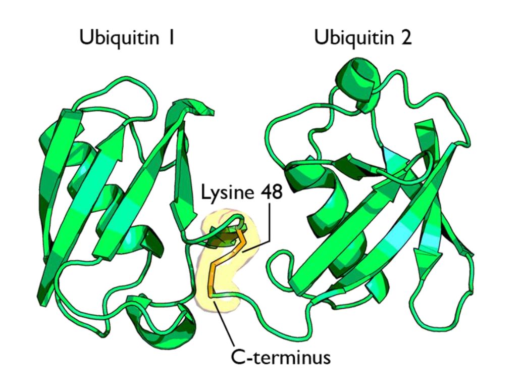
Fig 1 A cartoon representation of a lysine 48-linked diubiquitin molecule. The two ubiquitin chains are shown as green cartoons with each chain labelled. The components of the linkage are indicated and shown as orange sticks. By Rogerdodd (Own work) [CC BY-SA 3.0 (https://creativecommons.org/licenses/by-sa/3.0)], via Wikimedia Commons

The simplest type of chain is homogeneous (IOWs, ubiquitins are linked always at the same site). But many types of mixed and branched chains can also be found.

Let’s start with the most common situation: a poli-ubiquitination of (at least) 4 ubiqutins, linearly linked at K48. This is the common signal for proteasome degradation.

By the way, the 26S proteasome is another molecular machine of incredible complexity, made of more than 30 different proteins. However, its structure and function are not the object of this OP, and therefore I will not deal with them here.

The ubiquitin code is not completely understood, at present, but a few aspects have been well elucidated. Table 1 sums up the most important and well known modes:

Code

Meaning

Polyubiquitination (4 or more) with links at K48 or at K11 Proteasomal degradation
Monoubiqutination (single or multiple) Protein interactions, membrane trafficking, endocytosis
Polyubiquitination with links at K63 Endocytic trafficking, inflammation, translation, DNA repair.
Polyubiquitination with links at K63 (other links) Autophagic degradation of protein substrates
Polyubiquitination with links at K27, K29, K33 Non proteolytic processes
Rarer chain types (K6, K11) Under investigation


However, this is only a very partial approach. A recent bioinformatics paper:

An Interaction Landscape of Ubiquitin Signaling

(Paywall)

Has attempted for the first time a systematic approach to deciphering the whole code, using synthetic diubiquitins (all 8 possible variants) to identify the different interactors with those signals, and they identified, with two different methodologies,  111 and 53 selective interactors for linear polyUb chains, respectively. 46 of those interactors were identified by both methodologies.

The translation

But what “translates” the complex ubiquitin code, allowing ubiquinated proteins to met the right specific destiny? Again, we can refer to the diubiquitin paper quoted above.

How do cells decode this ubiquitin code into proper cellular responses? Recent studies have indicated that members of a protein family, ubiquitin-binding proteins (UBPs), mediate the recognition of ubiquitinated substrates. UBPs contain at least one of 20 ubiquitin-binding domains (UBDs) functioning as a signal adaptor to transmit the signal from ubiquitinated substrates to downstream effectors

But what are those “interactors” identified by the paper (at least 46 of them)? They are, indeed, complex proteins which recognize specific configurations of the “tag” (the ubiquitin chain), and link the tagged (ubiquinated) protein to other effector proteins which implement its final fate, or anyway contribute in deffrent forms to that final outcome.


The basic control of the procedure: the complexity of the ubiquitination process.

So, we have seen that ubiquitin chains work as tags, and that their coded signals are translated by specific interactors, so that the target protein may be linked to its final destiny, or contribute to the desired outcome. But we must still address one question: how is the ubiquitination of the different target proteins implemented? IOWs, what is the procedure that “writes” the specific codes associated to specific target proteins?

This is indeed the first step in the whole process. But it is also the most complex, and that’s why I have left it for the final part of the discussion.

Indeed, the ubiquitination process needs to realize the following aims:

  1. Identify the specific protein to be ubiquitinated
  2. Recognize the specific context in which that protein needs to be ubiquitinated
  3. Mark the target protein with the correct tag for the required fate or outcome

We have already seen that the ubiquitin system is involved in practically all different cellular paths and activities, and therefore we can expect that the implementation of the above functions must be a very complex thing.

And it is.

Now, we can certainly imagine that there are many different layers of regulation that may contribute to the general control of the procedure, specifically epigenetic levels, which are at present poorly understood. But there is one level that we can more easily explore and understand, and it is , as usual, the functional complexity of the proteins involved.

And, even at a first gross analysis, it is really easy to see that the functional complexity implied by this process is mind blowing.

Why? It is more than enough to consider the huge number of different proteins involved. Let’s see.

The ubiquitination process is well studied. It can be divided into three phases, each of which is implemented by a different kind of protein. The three steps, and the three kinds of proteins that implement them, take the name of E1, E2 and E3.
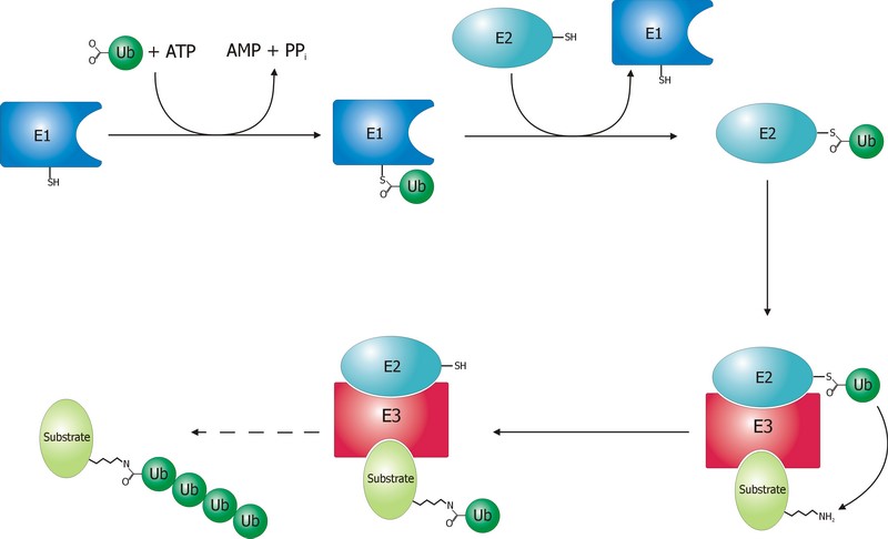
 

Fig. 2 Schematic diagram of the ubiquitylation system. Created by Roger B. Dodd: Rogerdodd at the English language Wikipedia [GFDL (http://www.gnu.org/copyleft/fdl.html) or CC-BY-SA-3.0 (http://creativecommons.org/licenses/by-sa/3.0/)], via Wikimedia Commons

 The E1 step of ubiquitination.

This is the first thing that happens, and it is also the simplest.

E1 is the process of activation of ubiquitin, and the E1 proteins is called E1 ubiquitin-activating enzyme. To put it simply, this enzyme “activates” the ubiquitin molecule in an ATP dependent process, preparing it for the following phases and attaching it to its active site cysteine residue. It is not really so simple, but for our purposes that can be enough.

This is a rather straightforward enzymatic reaction. In humans there are essentially two forms of E1 enzymes, UBA1 and UBA6, each of them about 1000 AAs long, and partially related at sequence level (42%).


The E2 step of ubiquitination.

The second step is ubiquitin conjugation. The activated ubiquitin is transferred from the E1 enzyme to the ubiquitin-conjugating enzyme, or E2 enzyme, where it is attached to a cysteine residue.

This apparently simple “transfer” is indeed a complex intermediate phase. Humans have about 40 different E2 molecules. The following paper:

E2 enzymes: more than just middle men

details some of the functional complexity existing at this level.

Abstract:

Ubiquitin-conjugating enzymes (E2s) are the central players in the trio of enzymes responsible for the attachment of ubiquitin (Ub) to cellular proteins. Humans have ∼40 E2s that are involved in the transfer of Ub or Ub-like (Ubl) proteins (e.g., SUMO and NEDD8). Although the majority of E2s are only twice the size of Ub, this remarkable family of enzymes performs a variety of functional roles. In this review, we summarize common functional and structural features that define unifying themes among E2s and highlight emerging concepts in the mechanism and regulation of E2s.

However, I will not go into details about these aspects, because we have better things to do: we still have to discuss the E3 phase!


The E3 step of ubiquitination.

This is the last phase of ubiquitination, where the ubiquitin tag is finally transferred to the target protein, as initial mono-ubiquitination, or to build an ubiquitin chain by following ubiqutination events. The proteins which implement this final passage are call E3 ubiquitin ligases. Here is the definition from Wikipedia:

A ubiquitin ligase (also called an E3 ubiquitin ligase) is a protein that recruits an E2 ubiquitin-conjugating enzyme that has been loaded with ubiquitin, recognizes a protein substrate, and assists or directly catalyzes the transfer of ubiquitin from the E2 to the protein substrate.

It is rather obvious that the role of the E3 protein is very important and delicate. Indeed it:

  1. Recognizes and links the E2-ubiquitin complex
  2. Recognizes and links some specific target protein
  3. Builds the appropriate tag for that protein (Monoubiquitination, mulptiple monoubiquitination, or poliubiquitination with the appropriate type of ubiquitin chain).
  4. And it does all those things at the right moment, in the right context, and for the right protein.

IOWs, the E3 protein writes the coded tag. It is, by all means, the central actor in our complex story.

So, here comes the really important point: how many different E3 ubiquitin ligases do we find in eukaryotic organisms? And the simple answer is: quite a lot!

Humans are supposed to have more than 600 different E3 ubiquitin ligases!

So, the human machinery for ubiquitination is about:

2 E1 proteins  –  40 E2 proteins – >600 E3 proteins

A real cascade of complexity!

OK, but even if we look at single celled eukaryotes we can already find an amazing level of complexity. In yeast, for example, we have:

1 or 2 E1 proteins  –  11 E2 proteins – 60-100 E3 proteins

See here:

The Ubiquitin–Proteasome System of Saccharomyces cerevisiae

Now, a very important point. Those 600+ E3 proteins that we find in humans are really different proteins. Of course, they have something in common: a specific domain.

From that point of view, they can be roughly classified in three groups according to the specific E3 domain:

  1. RING group: the RING finger domain ((Really Interesting New Gene) is a short domain of zinc finger type, usually 40 to 60 amino acids. This is the biggest group of E3s (about 600)
  2. HECT domain (homologous to the E6AP carboxyl terminus): this is a bigger domain (about 350 AAs). Located at the C terminus of the protein. It has a specific ligase activity, different from the RING   In humans we have approximately 30 proteins of this type.
  3. RBR domain (ring between ring fingers): this is a common domain (about 150 AAs) where two RING fingers are separated by a region called IBR, a cysteine-rich zinc finger. Only a subset of these proteins are E3 ligases, in humans we have about 12 of them.

See also here.

OK, so these proteins have one of these three domains in common, usually the RING domain. The function of the domain is specifically to interact with the E2-ubiquitin complex to implement the ligase activity. But the domain is only a part of the molecule, indeed a small part of it. E3 ligases are usually big proteins (hundreds, and up to thousands of AAs). Each of these proteins has a very specific non domain sequence, which is probably responsible for the most important part of the function: the recognition of the specific proteins that each E3 ligase processes.

This is a huge complexity, in terms of functional information at sequence level.

Our map of the ubiquinating system in humans could now be summarized as follows:

2 E1 proteins  –  40 E2 proteins – 600+ E3 proteins + thousands of specific substrates

IOWs, each of hundreds of different complex proteins recognizes its specific substrates, and marks them with a shared symbolic code based on uniquitin and its many possible chains. And the result of that process is that proteins are destined to degradation by the proteasome or other mechanisms, and that protein interactions and protein signaling are regulated and made possible, and that practically all cellular functions are allowed to flow correctly and smoothly.

Finally, here are two further compoments of the ubuquitination system, which I will barely mention, to avoid making this OP too long.

Ubiquitin like proteins (Ubl):

A number of ubiquitin like proteins add to the complexity of the system. Here is the abstract from a review:

The eukaryotic ubiquitin family encompasses nearly 20 proteins that are involved in the posttranslational modification of various macromolecules. The ubiquitin-like proteins (UBLs) that are part of this family adopt the β-grasp fold that is characteristic of its founding member ubiquitin (Ub). Although structurally related, UBLs regulate a strikingly diverse set of cellular processes, including nuclear transport, proteolysis, translation, autophagy, and antiviral pathways. New UBL substrates continue to be identified and further expand the functional diversity of UBL pathways in cellular homeostasis and physiology. Here, we review recent findings on such novel substrates, mechanisms, and functions of UBLs.

These proteins include SUMO, Nedd8, ISB15, and many others.

Deubiquitinating enzymes (DUBs):

The process of ubiquitination, complex as it already is, is additionally regulated by these enzymes which can cleave ubiquitin from proteins and other molecules. Doing so, they can reverse the effects of ubiquitination, creating a delicately balanced regulatory network. In humans there are nearly 100 DUB genes, which can be classified into two main classes: cysteine proteases and metalloproteases.


By the way, here is a beautiful animation of the basic working of the ubiquitin-proteasome system in degrading damaged proteins:


A summary:

So, let’s try a final graphic summary of the whole ubiquitin system in humans:
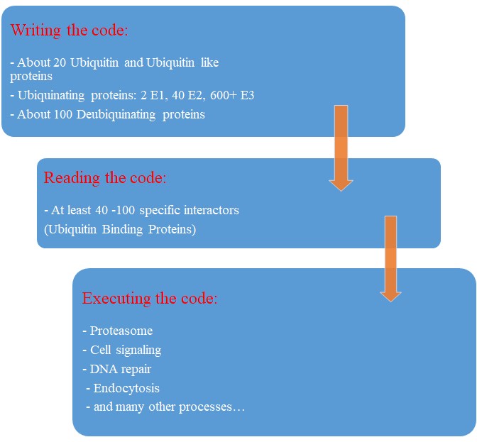
Fig 3 A graphic summary of the Ubiquitin System


Evolution of the Ubiquitin system?

The Ubiqutin system is essentially an eukaryotic tool. Of course, distant precursors for some of the main components have been “found” in prokaryotes. Here is the abstract from a paper that sums up what is known about the prokaryotic “origins” of the system:

Structure and evolution of ubiquitin and ubiquitin-related domains.

(Paywall)

Abstract:

Since its discovery over three decades ago, it has become abundantly clear that the ubiquitin (Ub) system is a quintessential feature of all aspects of eukaryotic biology. At the heart of the system lies the conjugation and deconjugation of Ub and Ub-like (Ubls) proteins to proteins or lipids drastically altering the biochemistry of the targeted molecules. In particular, it represents the primary mechanism by which protein stability is regulated in eukaryotes. Ub/Ubls are typified by the β-grasp fold (β-GF) that has additionally been recruited for a strikingly diverse range of biochemical functions. These include catalytic roles (e.g., NUDIX phosphohydrolases), scaffolding of iron-sulfur clusters, binding of RNA and other biomolecules such as co-factors, sulfur transfer in biosynthesis of diverse metabolites, and as mediators of key protein-protein interactions in practically every conceivable cellular context. In this chapter, we present a synthetic overview of the structure, evolution, and natural classification of Ub, Ubls, and other members of the β-GF. The β-GF appears to have differentiated into at least seven clades by the time of the last universal common ancestor of all extant organisms, encompassing much of the structural diversity observed in extant versions. The β-GF appears to have first emerged in the context of translation-related RNA-interactions and subsequently exploded to occupy various functional niches. Most biochemical diversification of the fold occurred in prokaryotes, with the eukaryotic phase of its evolution mainly marked by the expansion of the Ubl clade of the β-GF. Consequently, at least 70 distinct Ubl families are distributed across eukaryotes, of which nearly 20 families were already present in the eukaryotic common ancestor. These included multiple protein and one lipid conjugated forms and versions that functions as adapter domains in multimodule polypeptides. The early diversification of the Ubl families in eukaryotes played a major role in the emergence of characteristic eukaryotic cellular substructures and systems pertaining to nucleo-cytoplasmic compartmentalization, vesicular trafficking, lysosomal targeting, protein processing in the endoplasmic reticulum, and chromatin dynamics. Recent results from comparative genomics indicate that precursors of the eukaryotic Ub-system were already present in prokaryotes. The most basic versions are those combining an Ubl and an E1-like enzyme involved in metabolic pathways related to metallopterin, thiamine, cysteine, siderophore and perhaps modified base biosynthesis. Some of these versions also appear to have given rise to simple protein-tagging systems such as Sampylation in archaea and Urmylation in eukaryotes. However, other prokaryotic systems with Ubls of the YukD and other families, including one very close to Ub itself, developed additional elements that more closely resemble the eukaryotic state in possessing an E2, a RING-type E3, or both of these components. Additionally, prokaryotes have evolved conjugation systems that are independent of Ub ligases, such as the Pup system.


As usual, we are dealing here with distant similarities, but there is no doubt that the ubiquitin system as we know it appears in eukaryotes.

But what about its evolutionary history in eukaryotes?

We have already mentioned the extremely high conservation of ubiquitin itself.

UBA1, the main E1 enzyme, is rather well conserved from fungi to humans: 60% identity, 1282 bits, 1.21 bits per aminoacid (baa).

E2s are small enzymes, extremely conserved from fungi to humans: 86% identity, for example, for UB2D2, a 147 AAs molecule.

E3s, of course, are the most interesting issue. This big family of proteins behaves in different ways, consistently with its highly specific functions.

It is difficult to build a complete list of E3 proteins. I have downloaded from Uniprot a list of reviewed human proteins including “E3 ubiquitun ligase” in their name: a total of 223 proteins.

The mean evolutionary behavior of this group in metazoa is rather different from protein to protein. However, as a group these proteins exhibit an information jump in vertebrates which is significantly higher than the jump in all other proteins:
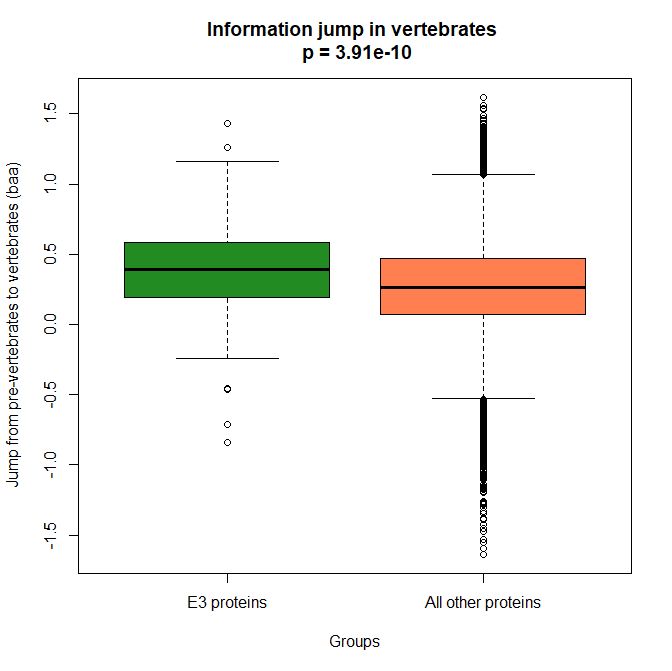
 

Fig. 4 Boxplots of the distribution of human conserved information jump from pre-vertebrates to vertebrates in 223 E3 ligase proteins and in all other human proteins. The difference is highly significant.


As we already know, this is evidence that this class of proteins is highly engineered in the transition to vertebrates. That is consistent with the need to finely regulate many cellular processes, most of which are certainly highly specific for different groups of organisms.

The highest vertebrate jump, in terms of bits per aminoacid, is shown in my group by the E3 ligase TRIM62. also known as DEAR1 (Q9BVG3), a 475 AAs long protein almost absent in pre-vertebrates (best hit 129 bits, 0.27 baa in Branchiostoma belcheri) and which flaunts an amazing jump of 1.433684 baa in cartilaginous fish (810 bits, 1.705263 baa).

But what is this protein? It is a master regulator tumor suppressor gene, implied in immunity, inflammation, tumor genesis.

See here:

TRIM Protein-Mediated Regulation of Inflammatory and Innate Immune Signaling and Its Association with Antiretroviral Activity

and here:

DEAR1 is a Chromosome 1p35 Tumor Suppressor and Master Regulator of TGFβ-Driven Epithelial-Mesenchymal Transition

This is just to show what a single E3 ligase can be involved in!

An opposite example, from the point of view of evolutionary history, is SIAH1, an E3 ligase implied in proteosomal degradation of proteins. It is a 282 AAs long protein, which already exhibits 1.787234 baa (504 bits) of homology in deuterostomes, indeed already 1.719858 baa in cnidaria. However, in fungi the best hit is only 50.8 bits (0.18 baa). So, this is a protein whose engineering takes place at the start of metazoa, and which exhibits only a minor further jump in vertebrates (0.29 baa), which brings the protein practically to its human form already in cartilaginous fish (280 identities out of 282, 99%). Practically a record.

So, we can see that E3 ligases are a good example of a class of proteins which perform different specific functions, and therefore exhibit different evolutionary histories: some, like TRIM62, are vertebrate quasi-novelties, others, like SIAH1, are metazoan quasi-novelties. And, of course, there are other behaviours, like for example BRCA1, Breast cancer type 1 susceptibility protein, a protein 1863 AAs long which only in mammals acquires part of its final sequence configuration in humans.

The following figure shows the evolutionary history of the three proteins mentioned above.
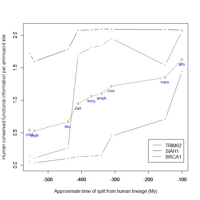
 

Fig. 5 Evolutionary history in metazoa of three E3 ligases (human conserved functional information)


An interesting example: NF-kB signaling

I will discuss briefly an example of how the Ubiquitin system interacts with some specific and complex final effector system. One of the best models for that is the NF-kB signaling.

NK-kB is a transcription factor family that is the final effector of a complex signaling pathway. I will rely mainly on the following recent free paper:

The Ubiquitination of NF-κB Subunits in the Control of Transcription

Here is the abstract:

Nuclear factor (NF)-κB has evolved as a latent, inducible family of transcription factors fundamental in the control of the inflammatory response. The transcription of hundreds of genes involved in inflammation and immune homeostasis require NF-κB, necessitating the need for its strict control. The inducible ubiquitination and proteasomal degradation of the cytoplasmic inhibitor of κB (IκB) proteins promotes the nuclear translocation and transcriptional activity of NF-κB. More recently, an additional role for ubiquitination in the regulation of NF-κB activity has been identified. In this case, the ubiquitination and degradation of the NF-κB subunits themselves plays a critical role in the termination of NF-κB activity and the associated transcriptional response. While there is still much to discover, a number of NF-κB ubiquitin ligases and deubiquitinases have now been identified which coordinate to regulate the NF-κB transcriptional response. This review will focus the regulation of NF-κB subunits by ubiquitination, the key regulatory components and their impact on NF-κB directed transcription.


The following figure sums up the main features of the canonical activation pathway:
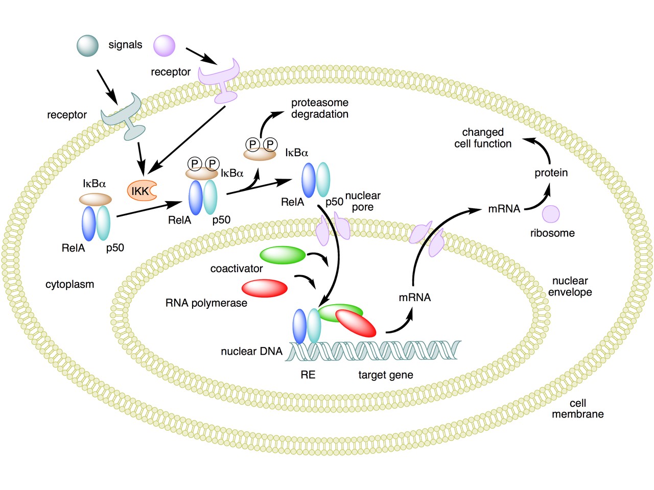
 

Fig. 6 A simple summary of the main steps in the canonical activayion pathway of NF-kB


Here the NF-κB TF is essentially the heterodimer RelA – p50. Before activation, the NF-κB (RelA – p50) dimer is kept in an inactive state and remains in the cytoplasm because it is linked to the IkB alpha protein, an inhibitor of its function.

Activation is mediated by a signal-receptor interaction, which starts the whole pathway. A lot of different signals can do that, adding to the complexity, but we will not discuss this part here.

As a consequence of receptor activation, another protein complex, IκB kinase (IKK), accomplishes the Phosphorylation of IκBα at serines 32 and 36. This is the signal for the ubiquitination of the IkB alpha inhibitor.

This ubiqutination targets IkB alpha for proteosomal degradation. But how is it achieved?

Well, things are not so simple. A whole protein complex is necessary, a complex which implements many different ubiquitinations in different contexts, including this one.

The complex is made by 3 basic proteins:

  • Cul1 (a scaffold protein, 776 AAs)
  • SKP1 (an adaptor protein, 163 AAs)
  • Rbx1 (a RING finger protein with E3 ligase activity, 108 AAs)

Plus:

  • An F-box protein (FBP) which changes in the different context, and confers specificity.

In our context, the F box protein is called beta TRC (605 AAs).
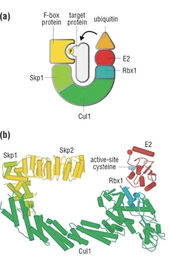
 

Fig. 7 A simple diagram of the SKP1 – beta TRC complex


Once the IkB alpha inhibitor is ubiquinated and degraded in the proteasome, the NF-κB dimer is free to translocate to the nucleus, and implement its function as a transcription factor (which is another complex issue, that we will not discuss).

OK, this is only the canonical activation of the pathway.

In the non canonical pathway (not shown in the figure) a different set of signals, receptors and activators acts on a different NF-κB dimer (RelB – p100). This dimer is not linked to any inhibitor, but is itself inactive in the cytoplasm. As a result of the signal, p100 is phosphorylated at serines 866 and 870. Again, this is the signal for ubiquitination.

This ubiquitination is performed by the same complex described above, but the result is different. P100 is only partially degraded in the proteasome, and is transformed into a smaller protein, p52, which remains linked to RelB. The RelB – p52 dimer is now an active NF-κB Transcription Factor, and it can relocate to the nucleus and act there.

But that’s not all.

  • You may remember that RelA (also called p 65) is one of the two components of NF-kB TF in the canonical pathway (the other being p 50). Well, RelA is heavily controlled by ubiquitination after it binds DNA in the nucleus to implement its TF activity. Ubiquitination (a very complex form of it) helps detachment of the TF from DNA, and its controlled degradation, avoiding sustained expression of NF-κB-dependent genes. For more details, see section 4 in the above quoted paper: “Ubiquitination of NF-κB”.
  • The activation of IKK in both the canonical and non canonical pathway after signal – receptor interaction is not so simple as depicted in Fig. 6. For more details, look at Fig. 1 in this paper: Ubiquitin Signaling in the NF-κB Pathway. You can see that, in the canonical pathway, the activation of IKK is mediated by many proteins, including TRAF2, TRAF6, TAK1, NEMO.
  • TRAF2 is a key regulator on many signaling pathways, including NF-kB. It is an E3 ubiquitin ligase. From Uniprot:  “Has E3 ubiquitin-protein ligase activity and promotes ‘Lys-63’-linked ubiquitination of target proteins, such as BIRC3, RIPK1 and TICAM1. Is an essential constituent of several E3 ubiquitin-protein ligase complexes, where it promotes the ubiquitination of target proteins by bringing them into contact with other E3 ubiquitin ligases.”
  • The same is true of TRAF6.
  • NEMO (NF-kappa-B essential modulator ) is also a key regulator. It is not an ubiquinating enzyme, but it is rather heavily regulated by ubiquitination. From Uniprot: “Regulatory subunit of the IKK core complex which phosphorylates inhibitors of NF-kappa-B thus leading to the dissociation of the inhibitor/NF-kappa-B complex and ultimately the degradation of the inhibitor. Its binding to scaffolding polyubiquitin seems to play a role in IKK activation by multiple signaling receptor pathways. However, the specific type of polyubiquitin recognized upon cell stimulation (either ‘Lys-63’-linked or linear polyubiquitin) and its functional importance is reported conflictingly.”
  • In the non canonical pathway, the activation of IKK alpha after signal – receptor interaction is mediated by other proteins, in particular one protein called NIK (see again Fig. 1 quoted above). Well, NIK is regulated by two different types of E3 ligases, with two different types of polyubiquitination:
    • cIAP E3 ligase inactivates it by constant degradation using a K48 chain
    • ZFP91 E3 ligase stabilizes it using a K63 chain

See here:

Non-canonical NF-κB signaling pathway.

In particular, Fig. 3

These are only some of the ways the ubiquitin system interacts with the very complex NF-kB signaling system. I hope that’s enough to show how two completely different and complex biological systems manage to cooperate by intricate multiple connections, and how the ubiquitin system can intervene at all levels of another process. What is true for the NF-kB signaling pathway is equally true for a lot of other biological systems, indeed for almost all basic cellular processes.

But this OP is already too long, and I have to stop here.

As usual, I want to close with a brief summary of the main points:

  1. The Ubiquitin system is a very important regulation network that shows two different signatures of design: amazing complexity and an articulated semiotic structure.
  2. The complexity is obvious at all levels of the network, but is especially amazing at the level of the hundreds of E3 ligases, that can recognize thousands of different substrates in different contexts.
  3. The semiosis is obvious in the Ubiquitin Code, a symbolic code of different ubiquitin configurations which serve as specific “tags” that point to different outcomes.
  4. The code is universally implemented and shared in eukaryotes, and allows control on almost all most important cellular processes.
  5. The code is written by the hundreds of E3 ligases. It is read by the many interactors with ubiquitin-binding domains (UBDs).
  6. The final outcome is of different types, including degradation, endocytosis, protein signaling, and so on.
  7. The interaction of the Ubiquitin System with other complex cellular pathways, like signaling pathways, is extremely complex and various, and happens at many different levels and by many different interacting proteins for each single pathway.

PS:

Thanks to DATCG for pointing to this video in three parts by Dr. Raymond Deshaies, was Professor of Biology at the California Institute of Technology and an Investigator of the Howard Hughes Medical Institute. On iBiology Youtube page:

A primer on the ubiquitin-proteasome system


Cullin-RING ubiquitin ligases: structure, structure, mechanism, and regulation


Targeting the ubiquitin-proteasome system in cancer

Comments
ET: Oh no! Not another corss-talk with TSZ! :) OK thanks for the notification. I checked TSZ and found the thread (rather short, for the moment, thanks God!). I must say that the paper you link was really mentioned by TomMueller, not Alan Fox (who is instead the author of the OP). Well, I am going to answer in next post. :)gpuccio
March 21, 2018
March
03
Mar
21
21
2018
12:26 AM
12
12
26
AM
PST
Alan Fox has taken umbrage with the claiming of ubiquitin as evidence for ID. Of course he doesn't have any idea how blind and mindless processes could have produced it and he doesn't have any idea how to test the claim that they could. So there. the alleged refutation of the concept of ubiquitin as evidence for ID Alan talks about alternatives- alternative to what? Alan's alleged evolutionary theory doesn't have some theory or hypothesis that could be a testable explanation for the ubiquitin system, to begin with.ET
March 20, 2018
March
03
Mar
20
20
2018
05:37 PM
5
05
37
PM
PST
DATCG: OK, I would like to go on with the more general discussion starting from what I have already said at #437. Why all the added complexity? I would like to start with an old friend, Michael Behe, and with one of his first metphors (in Darwin's black box): the Rude Goldberg machine. For example, here is an example: https://en.wikipedia.org/wiki/Rube_Goldberg_machine#/media/File:Rube_Goldberg%27s_%22Self-Operating_Napkin%22_(cropped).gif Now, it is rather obvious that the scenario we have seen at #437 about the NFkB pathway (see Fig. 1 of the quoted paper) does resemble a Rude Goldberg machine. But there is more. Behe offers, in his fundamental book, a couple of important examples of irreducible complexity: the bacterial flagellum and the coagulation cascade. But those examples are a little different from out regulation overkill. In a sense, the irreducible complexity is more "understandable" there. For example, in the flagellum the various parts, stator, rotor, filament and so on are parts of a machine. their role, therefore, is immediately obvious. In the coagulation cascade, the linear cascade can be explained, in a way, by the need to amplify linearly the signal to get a wide final effect. But in out regulation scenario, the explanation is less obvious. There is also another feature which can help us in our approach: the regulation network we have seen is not linear. If we look at the famous Fig. 1 already quoted, while the main cascade can be considered linear, there are many "cross-talks". For example, some phosphorylation systems and the ubiquitination step act "at the side" of the cascade. Now, I would propose what seems to me the only reasonable explanation for the "added complexity". The reason for the added complexity is that the pathway, like almost all similar pathways that work between the cell membrane and the nucleus, is not an isolated mechanism, but is part of a huge "neural network" which involves all the different pathways which transmit and integrate the communication between outward signals (the cell membrane) and the final transcription regulation in the nucleus. My idea is that the many proteins that are involved in the many "redundant" steps in the pathway are regulation nodes, and act as "sensors" which integrate the specific pathway with all that happens in the cytoplasm, receiving information from the other pathways and transmitting information to them. That is also an explanation to the more specific answer: Why are some apparently simple steps implemented by huge multi-protein complexes? For example, why is the double phosphorylation of IkB alpha performed by a structure made of 9 protein blocks, and not by a single kinase? One possible answer is: because it must receive and transmit information to other cell pathways, interacting with many other protein structures. IOWs, we see here something similar to what happens at the level of the nucleus with the combinatorial working of TFs in big multi-protein structures. Or, if we want to push the similitude even further, something similar to what happens in the synapses to integrate many different signals. OK, these are just a few tentative thoughts. But, if there is something true in these ideas, then the "neural network" of transmission pathways really deserved a lot of attention. And, of course, only a design perspective can help in this kind of issues.gpuccio
March 20, 2018
March
03
Mar
20
20
2018
02:50 PM
2
02
50
PM
PST
Note: @439 Bigger Picture: One question leads to another Why does a blind, unguided series of events "decide" in the past, millions of years ago to "evolve" a solution so that: a) a Pre-Formatted protein complex like NF-kB waits around in suspended animation for a cascade of events initiated by a signal event through a specific TNF pathway? Q: How many other Pre-formatted Protein Complexes are waiting in Suspended Animation of the Cytoplasm? The irreducible complexity of such networked systems amounts to multiple interdependent systems coordinating organized interactions while "simultaneously" and "blindly evolving" working solution steps at just the right time for any of these pathways to function. OK, now I must go. Will check in later. (Edit) and this does not include other possible pathways for this specific NF-kB protein complex for other factors(signals processing) and decisions. Either Pre or Post Phosphorylation and Ubiquitination.DATCG
March 19, 2018
March
03
Mar
19
19
2018
12:45 PM
12
12
45
PM
PST
Gpuccio, Before I go, restating for my own clarity and maybe others what you have stated re: "simpler" it might be on another pathway.
"...how simpler it would have been in this other way: 1) The interaction between TNF and its receptor enzumatically modifies a TF (p50-p65) OK, that's bypassing a huge Complex and many phosphorylation steps. So, am I correct in speculating Phosphorylation is crucial in ways I do not understand yet? 2) ...which was before in some inactive state. This is to me is an important Design concept or at least a good question: inactive state I question and expanded in previous comment @438. Why have NF-kB protein complex hanging around? I designate it as a "Pre-Formatted" state. Speed considerations? Quick response and deployment? Is there another Conditional element where different signals change the resulting pathway or proteolytic process? Why would a supposedly blind, unguided "process" do it? And how would it know it the Protein Complex would ever be utilized or "evolve" to be utilized in such a pathway? I've not seen previous remarks on this by neo-Darwinist. So if any readers supporting a neo-Darwinian pathway of evolution for this scenario can comment, I'd be happy to see it. Why does a blind, unguided series of events decide in the past, millions of years ago even that: a) I'm going to leave a Pre-Formatted protein complex like NF-kB waiting around in suspended animation? b) So a signal process can cause a chain reactions of Lemony Snicket series of "FORTUNATE" events? This does not make sense even for Lemony Snicket. 3) The active TF(edit) then relocates to the nucleus, where it does what it has to do. After a series of unfortunate Lemony Snicket's events! ;-) it proceeds to the Nucleus where a whole other series of unfortunate Lemony Snicket events translate, transcribe, Post-Modify and transpire to send an immune response to the target of the alert.
Amazing! :) That Lemony Snicket!
Simpler, isn’t it? And beware, that simpler version could easily be controlled too, for example at the level where the TF is activated by the membrane receptor. So, why all the added complexity?
Because, two kids showed up and created a new pathway for Lemony Snicket to go down, all by random walks, accidental events and "natural selection." .DATCG
March 19, 2018
March
03
Mar
19
19
2018
12:17 PM
12
12
17
PM
PST
Ciao Gpuccio @436-437 :) And thanks! Especially on clarifications and this one...
b) The phosphorylation described in a) is done by a big molecular complex, which includes the two IKK1 (or alpha) and IKK2 (or beta) proteins, NEMO and at least 3 other proteins (Hsp90, Cdc37 and ELKS). Many of those proteins must be phosphorylated to be active, in particular IKK1 at Ser 176 and 180, IKK2 at Ser 177 and 181.
To see IKK2(important role) at Ser 177 and 181 is interesting. I've missed something and thought it was Ser 32, Ser 36. Thanks for recognizing the question(however uninformed of biochemical pathways) about processes taking place - which seems like at initial look - to be over regulated for lack of better terminology. So, taking neo-Darwinist side, would a hodge-podge of pathways like this lead credence to their theory? Or, upon closer inspection will we find purposes for: a) multiple pathways b) reasons for phosphorylation steps c) ubiquitinylatoin steps d) Reason for Pre-Formatted NF-kB Waiting in the Cytoplasm? I'd really like a neo-Darwinist to explain D) Pre-formatted TF w/ inhibitor awaiting signals. Why? Why is is there? Makes no since for an unguided, blind process to pre-plan actions. Does it? Will neo-Darwinist appeal to bad design as an answer? So yes, the question again, is Why? Why for many of these steps. I was in the middle of breaking down the two pathways of Figure 1, Classical vs Alternative(TNF vs CD40) and simply do not have enough time at this moment. But will hopefully return later tonight and finish before posting. But what we see is Directed Response pathways for TNF and CD40, plus a third pathway which I'm not planning to cover. I like how you are detailing what might happen in a simpler pathway - if only we knew all of these processing techniques and other components. I think the more we peer in to different Signaling actions and Pathways, starting at the membrane, the more beneficiary it is to a Design hypothesis. First, it must distinguish between TNF and CD40. These are conditional signals for appropriate specified and targeted immune responses(ie. viruses in alternative pathway of CD40) OK, have very busy week ahead, but I'll respond when I can! Really enjoying this OP Gpuccio! And I hope other readers appreciate all your efforts here. And hope they feel OK to ask any questions.DATCG
March 19, 2018
March
03
Mar
19
19
2018
11:58 AM
11
11
58
AM
PST
DATCG at #432: Now, the more general discussion, for which I will use our NFkB as a model, but there are lots of similar systems in the cell. In your comment you have touched a very important point. An extremely important point, I would say. In brief, I will sum it up as followes: Why such complex multiple intertwined regulation systems to control one single pathway? I suggest that anyone who reads the following could refer to Fig. 1 in the already quoted paper: https://orbi.uliege.be/bitstream/2268/1280/1/21.%20Review%20phosphorylation%20NF-kB%20TIBS.pdf That will make the discussion simpler. Please, refer only to the left part of the Figure, which represents the canonical activation pathway. Now, the whole process coule be summarized as follows: a) A signal arrives at the cell membrane (The TNF cytokine) b) A transcription factor is activated to convey the message to the nucleus. c) The activated TF interacts with the genome and aucses a series of events there. In our Fig. 1, it is easy to identify the eseential actors. a) The cytokine (TNF) and its membrane receptor (TNFR1) are well visible at the top of the Figure (left part). b) Our TF is the p50-p65 entity (p65 is the same as RelA, so this is the same as RelA-p50 in Fig. 5 of the OP). c) The TF interacting with DNA can be seen at the bottom of the Figure. OK, so I think that the spontaneous question that everyone is asking is: But there are a lot of other things there! Why? And there are! a) The protein complex which forms at the inner side of the cell membrane (3 proteins, one of which phosphorylated, + SODD which has to be released for the activation) b) The big protein complex which phosphorylates the TF to release it from its inhibitor: 6 proteins, many of which phosphorylated. c) The inhibitor of the TF (IkB alpha) d) The SKP1 – beta TRC complex which ubiquinates the inhibitor after its double phosphorylation (not shown in the figure, see Fig. 7 in the OP): 6 proteins, including ubiquitin. e) And, of course, the proteasome. And I would bet that a lot of other components are not shown, maybe even not known (for example, the various Kinase systems that phosphorylate many of the mentioned proteins). Each of those components has an active role in the regulation and control of the process. And again, the question is: why? Of course, anyone can see how simpler it would have been in this other way: The interaction between TNF and its receptor enzumatically modifies a TF (p50-p65) which was before in some inactive state. The active TF then relocates to the nucleus, where it does what it has to do. Simpler, isn't it? And beware, that simpler version could easily be controlled too, for example at the level where the TF is activated by the membrane receptor. So, why all the added complexity? That is a difficult question. I will try to discuss some aspects in next post.gpuccio
March 19, 2018
March
03
Mar
19
19
2018
10:09 AM
10
10
09
AM
PST
DATCG: First of all, I would like to offer some clarifications about the complex issue of phosphorylation in the NFkB pathway, just to avoid confusion. a) The protein which is phosphorylated at serines 32 and 36 is IkB alpha, which is a direct inhibitor of RelA p50 (in the canonocal pathway of activation). The double phopshorylation leads to ubiquitination of the inhibitor and to its degradation in the proteasome, releasing the transcription factor (RelA p50, which is one form of NFkB TF). b) The phosphorylation described in a) is done by a big molecular complex, which includes the two IKK1 (or alpha) and IKK2 (or beta) proteins, NEMO and at least 3 other proteins (Hsp90, Cdc37 and ELKS). Many of those proteins must be phosphorylated to be active, in particular IKK1 at Ser 176 and 180, IKK2 at Ser 177 and 181. c) Not much is understood of the processes that lead to the phosphorylations described in b). The TAK1 kinase seems to be involved. d) However, the formation of the protein complex described in b) is cuased by another protein complex, which adheres to the cell membrane, and includes TRADD, TRAF2 and RIP (this one, too, phosphorylated). e) The formation of the complex described in d) is caused by the reaction between a specific membrane receptor and a cytokine (usually Tumor Necrosis Factor, TNF). Simple, isn't it? And this is only the canonical pathway! :) OK, the above information is not extremely recent, it comes from the following two papers: Phosphorylation of NF-kB and IkB proteins: implications in cancer and inflammation https://orbi.uliege.be/bitstream/2268/1280/1/21.%20Review%20phosphorylation%20NF-kB%20TIBS.pdf See especially Fig. 1. And: The IKK Complex, a Central Regulator of NF-?B Activation https://www.ncbi.nlm.nih.gov/pmc/articles/PMC2829958/pdf/cshperspect-NFK-a000158.pdf I just wanted to clarify these points to go on with some more general discussion in next post.gpuccio
March 19, 2018
March
03
Mar
19
19
2018
07:00 AM
7
07
00
AM
PST
So, is that activity around serines 177 and 181 applicatlbe to serines 32 and 36? I don't know if same rules apply. It is from another report in the book linked in #434, that describes... "two potentially novel components of the IKK complex, namely Cdc37 and Hsp90. Apparently, formation of the core IKK complex with Cdc37/Hsp90 is required for TNF induced activation and recruitment of the core IKK complex from the Cytoplasm to the membrane." Different functional requirements may be unrelated.DATCG
March 18, 2018
March
03
Mar
18
18
2018
06:46 PM
6
06
46
PM
PST
Hmmmm... "Phosphorylation of IkBalpha on serines 32 and 36 is mediated by IkB kinases(IKKs), whose activity is induced by activators of the NFkB pathway. IKK activity exists as large Cytoplasmic multi-subunit complex(700-900kDAa) containing two kinase subunits, IKK1(IKKalpha) and IKK2(IKKbeta), and regulartory subunit, NEMO..." "Sequence analysis revealed that both IKK1 and IKK2 contain a canonical MAP kinase(MAPKK) activation loop motif. This region contains specific sites whose phosphorylation induces a conformational change that results in kinase activation." Hmmm, is that Kinase activation in support of Tagging. OK, so this is interesting. Paragraphs above and blockquote below are from google book source. There may be a few typos: Regulation of Organelle and Cell Compartment Signaling: Cell Signaling... edited by Ralph A. Bradshaw, Edward A. Dennis(1st Edition 2011).
Phosphorylation within the activation loop typically occurs through the action of an upstream kinase or through transphosphorylation enabled by regulated proximity between two kinase subunits. IKK2 activation loop mutations, in which serines 177 and 181 were replaced with alanine, render the kinase refarctory to stimulus dependent activiatio. In contrast, replacement of serine 177 and 181 with glutamic acid, to mimic phoserine, yielded a constitutively active kinase, and was capable of cell stimulation. The corresponding mutations in IKK1 did not interfere wiht NFkB activation in response to IL-1 or TNF, providing the first data suggesting that IKK2 plays a more prominent role in NFkB activation in response to proinflammatory cytyokines.
.DATCG
March 18, 2018
March
03
Mar
18
18
2018
06:19 PM
6
06
19
PM
PST
Correction of sentence from 432 above: It must match the signal input. And in turn the pathway has to launch the correct response by a Directive or Rules based procedure code that has addresses or addressable-location mechanisms built-in to guide for eventual locations and active response(i.e. inflammatory and/or immune responses must be directed to correct area of inflammation and repair)DATCG
March 18, 2018
March
03
Mar
18
18
2018
05:37 PM
5
05
37
PM
PST
Hey guys, hope everyone had a great weekend, restful and invigorating :) Gpuccio @425 - 430! :) Wow! It's always fun to read all the spectacular details of what you've posted. I especially like Splicesome Ubiquitin connections as expected. And I'd love to delve into that area you highlighted, especially a favorite area of introns for me :) My time is limited however so will focus on the part of your OP on Phosphorylation --------------------- On #425... building upon an analogy of Design process and Markup Languages... The Phosphorylation Tag as a refined, or fine tuning condition step is very interesting. What is it accomplishing and why precede UPS with a unique tag? Whats the reason for this specific pathway? To release a transcription factor? For that matter, why is this a Pre-release state required as a partial degradation for release? Weird, huh? Is there a precedent in Design and Coding realms for an Information processing analogy? There's a reason Bill Gates said, “DNA is like a computer program but far, far more advanced than any software ever created.” Well, why would he say that? Because he is intimately aware of how a CPU works with Coding languages. He recognizes Code when he sees it in an operating system. So yes, there is a precedent in information processing. Programmers maintain internal information, code, data and append internal tags or external tags in a table or database and strip data or "inhibitors" if you like as Pre-processing requirements are met, based upon different input signals. So, if we view the Cytoplasm as another method of large CPU-processing Memory in the Cell, Or, lets call it an "aqueous MotherBoard*" this begins to make sense as an Information Processing analogy. Data or in this case, the NF-?B TF Struture is Pre-formatted. That alone is a sign of Design. The NF-kB Transcription Factor is ready and waiting for activation to instigate it's release based upon specific input signal(s) for it's eventual modification and release into the Nucleus. One of my first questions is... Why have a pre-formatted structure waiting for use in a blind, unguided neo-Darwinian story? This Pre-formatted TF does not fire off for any reason. In fact, if it starts firing off for any reason, it would cause chaos and possible damage downstream. So it's tightly regulated and only released upon correct Signal(s). It must match the signal input. And in turn the pathway has to launch the correct response by a Directive or Rules based procedure code that has locations built-in for eventual locations. It's like a data-table or structure is be preloaded into memory ready for quick access and retrieval by a CPU-nucleus instruction set for transcription. This is not a single Input -> Modification -> Output process. These rapid process are going on in parallel, especially for quick reaction immune systems and/or repair mechanisms and any millions of trillions of cellular transactions at any one time in our body, brain, heart, skin, immune, gut, etc. etc., etc. But simplifying, we have... Input to Aqueous Cytoplasm "Motherboard" for the Release of Transcription Factor : -> Signal A ---> Structure Awaiting Signal A in Wet Memory -----> Tag for Modification -------> Alert UPS ---------> Strip Component Part, freeing TF -----------> Send NF-?B TF to Nucleus for processing An interesting question arises which I think is even more succinct and descriptive of a Design process. Why not ubiquitin Mono, Poly, or Branched tags? Why a requirement for a conditional, two-step Tagging process? Why not a one-step UPS solution or chaining events? That would satisfy partial degradation and release of TF to the nucleus? I need more time to review. Unfortunately I do not have enough background in biochemistry and atomic structures to know why these different Tagging procedures might be required. A few searches have not produced answers though I may easily be missing them. Is it a conformational component where Phosphorylation is required to adjust folds? If by Design, we should be able to qualify the reasons for conditions of refinement specificity that you mention and Tagging requirements. So much to review :) *Aqueous Motherboard - what else can we call it? Or designate the Cytoplasm as? Other than it's aqueous structure of floating functions and organelles - it resembles a motherboard. The Cytoplasm functions as a motherboard for active, instant retrieval of Pre-formatted structures, specified functions and specialized processing units(1). These units and floating pre-formatted functions surround the CPU - Core process-Nucleus instruction set for Eukaryotes. This enables high speed throughput, as signals enter and ignite pre-programmed responses. (1) specialized-processing units = organelles. What am I missing? What more might be added I'm leaving out in symbolic tagging and semiosis? Or processing functionality?DATCG
March 18, 2018
March
03
Mar
18
18
2018
05:08 PM
5
05
08
PM
PST
UB "ES, I do not see any mail, my friend". That explains your silence :) I'll send another one. Thanks for letting me know. I always enjoy reading this kind of OPs, as you know. But time is really tight just now. I hope to be able to devote more time to it in due course.EugeneS
March 18, 2018
March
03
Mar
18
18
2018
01:39 PM
1
01
39
PM
PST
DATCG: Here is another protein regulated by ubiquitin and involved in splicing, Sde2: Sde2 is an intron-specific pre-mRNA splicing regulator activated by ubiquitin-like processing. http://emboj.embopress.org/content/37/1/89.long
Abstract The expression of intron?containing genes in eukaryotes requires generation of protein?coding messenger RNAs (mRNAs) via RNA splicing, whereby the spliceosome removes non?coding introns from pre?mRNAs and joins exons. Spliceosomes must ensure accurate removal of highly diverse introns. We show that Sde2 is a ubiquitin?fold?containing splicing regulator that supports splicing of selected pre?mRNAs in an intron?specific manner in Schizosaccharomyces pombe. Both fission yeast and human Sde2 are translated as inactive precursor proteins harbouring the ubiquitin?fold domain linked through an invariant GGKGG motif to a C?terminal domain (referred to as Sde2?C). Precursor processing after the first di?glycine motif by the ubiquitin?specific proteases Ubp5 and Ubp15 generates a short?lived activated Sde2?C fragment with an N?terminal lysine residue, which subsequently gets incorporated into spliceosomes. Absence of Sde2 or defects in Sde2 activation both result in inefficient excision of selected introns from a subset of pre?mRNAs. Sde2 facilitates spliceosomal association of Cactin/Cay1, with a functional link between Sde2 and Cactin further supported by genetic interactions and pre?mRNA splicing assays. These findings suggest that ubiquitin?like processing of Sde2 into a short?lived activated form may function as a checkpoint to ensure proper splicing of certain pre?mRNAs in fission yeast.
And: Intron specificity in pre-mRNA splicing https://link.springer.com/article/10.1007%2Fs00294-017-0802-8
Abstract The occurrence of spliceosomal introns in eukaryotic genomes is highly diverse and ranges from few introns in an organism to multiple introns per gene. Introns vary with respect to their lengths, strengths of splicing signals, and position in resident genes. Higher intronic density and diversity in genetically complex organisms relies on increased efficiency and accuracy of spliceosomes for pre-mRNA splicing. Since intron diversity is critical for functions in RNA stability, regulation of gene expression and alternative splicing, RNA-binding proteins, spliceosomal regulatory factors and post-translational modifications of splicing factors ought to make the splicing process intron-specific. We recently reported function and regulation of a ubiquitin fold harboring splicing regulator, Sde2, which following activation by ubiquitin-specific proteases facilitates excision of selected introns from a subset of multi-intronic genes in Schizosaccharomyces pombe
Both of January 2018. Strangely, Sde2 is not linked to splicing in Uniprot:
Involved in both DNA replication and cell cycle control (PubMed:27906959). Unprocessed SDE2 interacts with PCNA via its PIP-box. The interaction with PCNA prevents monoubiquitination of the latter thereby inhibiting translesion DNA synthesis. The binding of SDE2 to PCNA also leads to processing of SDE2 by an unidentified deubiquinating enzyme, cleaving off the N-terminal ubiquitin-like domain. The resulting mature SDE2 is degraded by the DCX(DTL) complex in a cell cycle- and DNA damage dependent manner (PubMed:27906959). Binding of SDE2 to PCNA is necessary to counteract damage due to ultraviolet light induced replication stress. The complete degradation of SDE2 is necessary to allow S-phase progression
So, its role in intron splicing seems to be a really recent discovery. By the way, human Sde2 is not highly conserved, but has a rather slow emergence in evolutionary history, with two dicrete jumps at the vertebrate and mammal transitions.gpuccio
March 18, 2018
March
03
Mar
18
18
2018
12:55 PM
12
12
55
PM
PST
DATCG: Well, the issue is more complex than I thought. It seems that NineTeen complex is involved in the many phases of the spliceosome assembly, with all the activities described in the paper linked at #428. However, the complex itself is formed by at least 8 core proteins (in yeats): Prp19, Cef1, Syf1, Syf2, Syf3, Snt309, Isy1 and Ntc20, plus about 18 associated proteins. See here: The function of the NineTeen Complex (NTC) in regulating spliceosome conformations and fidelity during pre-mRNA splicing. https://www.ncbi.nlm.nih.gov/pmc/articles/PMC4234902/
Abstract: The NineTeen Complex (NTC) of proteins associates with the spliceosome during pre-mRNA splicing and is essential for both steps of intron removal. The NTC and other NTC-associated proteins are recruited to the spliceosome where they participate in regulating the formation and progression of essential spliceosome conformations required for the two steps of splicing. It is now clear that the NTC is an integral component of active spliceosomes from yeast to humans and provides essential support for the spliceosomal snRNPs (small nuclear ribonucleoproteins). In the present article, we discuss the identification and characterization of the yeast NTC and review recent work in yeast that supports the essential role for this complex in the regulation and fidelity of splicing.
In particular, Table 1 But the strange point is that:
The NTC is named after the splicing factor Prp19, which was first identified in 1993 as a splicing factor in the yeast S. cerevisiae [4]. Prp19 is essential for splicing but is not a constituent of any of the individual spliceosomal snRNPs [5,6]. Association of Prp19 with itself to form tetramers provides the basis for the hypothesis that Prp19 provides a scaffold for NTC organisation [7]. Prp19 contains a U-box domain which exhibits E3 ubiquitin ligase activity in vitro [8], however, a target for this activity in the spliceosome is still lacking.
Emphasis mine. IOWs, Prp19 is definitely an E3 ligase, but that activity is not documented in the spliceosome assembly (although it could certainly be present). However, the very recent paper referenced at #427 clearly documents the E3 ligase activity, but in relation to DNA repair, not to spliceosome assembly. Moreover, the 3 proteins listed at #427 are required for the E3 ligase activity in DNA repair, but are not apparently part of the NTC complex, as I had believed initially (I have corrected my comment in that sense). These proteins are really amazing, I would say. :)gpuccio
March 18, 2018
March
03
Mar
18
18
2018
12:16 PM
12
12
16
PM
PST
DATCG: And this is about the NineTeen Complex (NTC): The NineTeen Complex (NTC) and NTC-associated proteins as targets for spliceosomal ATPase action during pre-mRNA splicing. https://www.ncbi.nlm.nih.gov/pmc/articles/PMC4615276/
Abstract: Pre-mRNA splicing is an essential step in gene expression that removes intron sequences efficiently and accurately to produce a mature mRNA for translation. It is the large and dynamic RNA-protein complex called the spliceosome that catalyzes intron removal. To carry out splicing the spliceosome not only needs to assemble correctly with the pre-mRNA but the spliceosome requires extensive remodelling of its RNA and protein components to execute the 2 steps of intron removal. Spliceosome remodelling is achieved through the action of ATPases that target both RNA and proteins to produce spliceosome conformations competent for each step of spliceosome activation, catalysis and disassembly. An increasing amount of research has pointed to the spliceosome associated NineTeen Complex (NTC) of proteins as targets for the action of a number of the spliceosomal ATPases during spliceosome remodelling. In this point-of-view article we present the latest findings on the changes in the NTC that occur following ATPase action that are required for spliceosome activation, catalysis and disassembly. We proposed that the NTC is one of the main targets of ATPase action during spliceosome remodelling required for pre-mRNA splicing.
Look at Fig. 1 for a "simple" summary. :)gpuccio
March 18, 2018
March
03
Mar
18
18
2018
11:57 AM
11
11
57
AM
PST
DATCG: OK, it had to happen: Here are the ubiquitin system and the spliceosome joined together, with additional involvement in DNA repair. And in a very complex way. Mol Cell. 2018 Mar 15: Prp19/Pso4 Is an Autoinhibited Ubiquitin Ligase Activated by Stepwise Assembly of Three Splicing Factors
Abstract Human nineteen complex (NTC) acts as a multimeric E3 ubiquitin ligase in DNA repair and splicing. The transfer of ubiquitin is mediated by Prp19-a homotetrameric component of NTC whose elongated coiled coils serve as an assembly axis for two other proteins called SPF27 and CDC5L. We find that Prp19 is inactive on its own and have elucidated the structural basis of its autoinhibition by crystallography and mutational analysis. Formation of the NTC core by stepwise assembly of SPF27, CDC5L, and PLRG1 onto the Prp19 tetramer enables ubiquitin ligation. Protein-protein crosslinking of NTC, functional assays in vitro, and assessment of its role in DNA damage response provide mechanistic insight into the organization of the NTC core and the communication between PLRG1 and Prp19 that enables E3 activity. This reveals a unique mode of regulation for a complex E3 ligase and advances understanding of its dynamics in various cellular pathways. --- The Prp19/nineteen complex (NTC) is a multifunctional protein complex involved in very diverse biological processes, including pre-mRNA splicing and the DNA damage response (DDR)
Prp19 is one more Prp involved, among other things, in the spliceosome assembly. From Uniprot:
Ubiquitin-protein ligase which is a core component of several complexes mainly involved pre-mRNA splicing and DNA repair. Core component of the PRP19C/Prp19 complex/NTC/Nineteen complex which is part of the spliceosome and participates in its assembly, its remodeling and is required for its activity. During assembly of the spliceosome, mediates 'Lys-63'-linked polyubiquitination of the U4 spliceosomal protein PRPF3. Ubiquitination of PRPF3 allows its recognition by the U5 component PRPF8 and stabilizes the U4/U5/U6 tri-snRNP spliceosomal complex (PubMed:20595234). Recruited to RNA polymerase II C-terminal domain (CTD) and the pre-mRNA, it may also couple the transcriptional and spliceosomal machineries (PubMed:21536736). The XAB2 complex, which contains PRPF19, is also involved in pre-mRNA splicing, transcription and transcription-coupled repair (PubMed:17981804). Beside its role in pre-mRNA splicing PRPF19, as part of the PRP19-CDC5L complex, plays a role in the DNA damage response/DDR. It is recruited to the sites of DNA damage by the RPA complex where PRPF19 directly ubiquitinates RPA1 and RPA2. 'Lys-63'-linked polyubiquitination of the RPA complex allows the recruitment of the ATR-ATRIP complex and the activation of ATR, a master regulator of the DNA damage response (PubMed:24332808). May also play a role in DNA double-strand break (DSB) repair by recruiting the repair factor SETMAR to altered DNA (PubMed:18263876). As part of the PSO4 complex may also be involved in the DNA interstrand cross-links/ICLs repair process (PubMed:16223718). In addition, may also mediate 'Lys-48'-linked polyubiquitination of substrates and play a role in proteasomal degradation (PubMed:11435423). May play a role in the biogenesis of lipid droplets (By similarity). May play a role in neural differentiation possibly through its function as part of the spliceosome
So many proteins involved in this extraordinary multi-protein complex: Prp19 (504 AAs) (as an homotetramer) SPF27 (225 AAs) CDC5L (802 AAs) PLRG1 (514 AAs) All of them highly conserved! :)gpuccio
March 18, 2018
March
03
Mar
18
18
2018
11:51 AM
11
11
51
AM
PST
DATCG: By the way, have you seen that our private party here has got a lot of new visibility, thanks to this kind OP by Barry Arrington? :) https://uncommondescent.com/intelligent-design/they-wont-dance-they-wont-mourn/gpuccio
March 18, 2018
March
03
Mar
18
18
2018
04:42 AM
4
04
42
AM
PST
DATCG at #421: "This Ubiquitin post of yours sparked my interest more than usual." Mine too, I must say. "Precisely because it’s a Tagging system. Or Markup Language as an analogy?" Exactly. The tagging/markup analogy is perfectly justified! I would like to add a few thoughts about the nature of the tag. Of course, ubiquitin is not the only "tag" here. We have seen that many other systems cooperate, and some of them just give the appropriate signal to the ubiquitin system. One good example is phosphorylation, which often serves as a tag to recruit the ubiquitin system to some specific target. So, we have a double specificity here: the ubiquitin system recognizes the target (usually by the E3 ligase), and it also recognizes the tag (phosphorylation). A good example of that mechanism can be found in the OP, where it is mentioned that phosphorylation of I?B? at serines 32 and 36 is the signal for the ubiquitination of the IkB alpha inhibitor (See Fig. 6). OK, so what is the main difference between, say, the phosphorylation tag and the ubiquitin tag? I would say that it is the fact that ubiquitin is a collection of different tags: IOWs, the system is much richer. Phosphorylation is a very powerful tag, but it is one tag, and therefore its symbolic meaning is linked essentially to the positions that are phosporylated in the target porteoin. For example, serines 32 and 36 inb the case of I?B?. The same would be true for ubiquitin if only mono-ubiquitination, single or multiple, existed. Then we would have one tag, whcih can assume different meanings according to the positions where it is added. But, as we well know, things are much more complex for ubiquitin. Much of the signaling, here, is made not ny mono-ubiquitination, but by ubiquitin chains. So, while the position where the chian is added retains all its symbolic meaning, a new layer of coding is added: the length and nature of the chain. In that sense, ubiquitin is really a miraculous peotein. Its special fold provides 8 different switches that can be used to build chains. So we have the following combinatorial degrees of freedom: a) The length of the chain can vary b) Homogeneous chains can be buith using each of the possible switches. c) Heterogeneous chains can be built by mixing different switches. That's simply outstanding! :)gpuccio
March 18, 2018
March
03
Mar
18
18
2018
04:40 AM
4
04
40
AM
PST
ES, I do not see any mail, my friend.Upright BiPed
March 18, 2018
March
03
Mar
18
18
2018
12:12 AM
12
12
12
AM
PST
DATCG at #421: Wow! What a tour the force! As usual, it is late now. I will comment on it tomorrow! :)gpuccio
March 17, 2018
March
03
Mar
17
17
2018
04:19 PM
4
04
19
PM
PST
EugeneS: Nice to see you here! :) I hope you will like the discussion about our friend ubiquitin. It has been much wider than each of us expected. And, of course, any comments from you will be greatly appreciated! :)gpuccio
March 17, 2018
March
03
Mar
17
17
2018
04:17 PM
4
04
17
PM
PST
Upright Biped, Gpuccio, Thanks guys, enjoy learning from you both! Gpuccio, your background in medicine helps so much. And I like your detailed analysis. This Ubiquitin post of yours sparked my interest more than usual. Precisely because it's a Tagging system. Or Markup Language as an analogy? Most of what I worked on dealt with conditional processing of language specific identifiers, imaging systems, document management and packaging. Input Processing, Tagging and Rules based systems were created to coordinate a tightly controlled decision tree of subroutines. Built on specific language requirements used across all 50 states. All of it controlled by municipalities, medical boards, state and federal regulations. Lots of legalese w/ medical and beneficiary enrollment plans - healthcare. For large corporations and government it was a lengthy process and a possible legal nightmare if a single mistake(mutation) was made. Every decision made revolved around Tagging and Rules Based language procedures for Identification and Information processing routines. It was seen as fairly revolutionary at the time by a small software startup. What traditionally took six months to a year, even two years in large cases were reduced to mere days or weeks(and that only due to Human reviews). Simpler applications reduced it to mere seconds for request to end users. We were doing Markup Language and Tagging before HTML was fully accepted. Looking back realized I worked with some of the brightest in the industry. Visionary developers at that time. Was a great experience. These were legacy systems that eventually crossed over to PC and Browsers. Fortunately, I was chosen to bridge the gap for a few clients. So I learned many different platforms and markup languages over the years. OK, all this to explain my interest in this post Gpuccio. Going back to your OP, you identified Ubiquitin as a Tagging Solution. Precisely! In my view this Tagging solution and "Markup Language" or Ubiquitin Code Identification leaped out as Design from the start. Functional information processing systems or Cellular Processing cannot exist without Identification and Tagging or Marking Codes. A quick review of your initial post(note: edited)
The semiosis: the ubiquitin code The title of this OP makes explicit reference to semiosis. Let’s try to see why. The simplest way to say it is: ubiquitin is a tag. The addition of ubiquitin to a substrate protein marks that protein for specific fates, the most common being degradation by the proteasome. Nonproteolytic Functions of Ubiquitin in Cell Signaling
Abstract: In the past few years..., nonproteolytic functions of ubiquitin have been uncovered at a rapid pace. These functions include <b<(Tagging of:) membrane trafficking, protein kinase activation, DNA repair, and chromatin dynamics. A common mechanism(Tagging) underlying these functions is that ubiquitin, or polyubiquitin chains, serves as a signal to recruit proteins harboring ubiquitin-binding domains, thereby bringing together ubiquitinated proteins and ubiquitin receptors to execute specific biological functions. Another important aspect is that ubiquitin is not one tag, but rather a collection of different tags. IOWs, a tag based code.
Bingo :) a "Tag Based Code" Or, Markup Language Identifier? One area of clarification. Gpuccio you and Dionisio previously highlighted and discussed the missing procedures? Where are the governing Rules and procedures? Thinking from a Design interpretation. If we look at it through combination of instantiated information processing(substrates, Tags and Markings), we see a series of different Markup Languages and Tagging Identifiers. Mono, Poly-ubiquitin and branch Ubiquitin, etc. Can we designate these as external Tags(markups)? Based upon an information tagging processes being researched and discovered today of regulatory UB systems. But, what is missing? Might it be an internal Rules based, Tagging system? Back to Language Markup principles and Design. We used internal Markup Languages and Rules for identification of external tag markers, conditions based or Contextual and systems based, including transformation across different coded networks and languages. We internally marked(Tagged) every bit of language in whole document pagkages by Document IDs, pages, sections, paragraphs, down to single words, even characters, and internal translations, including special post-processing moditifications. All of this modular packaging sytems clients could pick and choose for whatever best represented their requirements. There was external Markup Language that End Users edited documents with, as identifiers to internal systems processing and tagging for eventual output destinations and other decision-required processing. All of it regulated by internal identifiers - Tags and Procedures only Developers could change or update. Overtime we allowed more overrides by customers to speed-up client specializations and less dependency on Developers. A more Open based User friendly markup. Q: Are we are on a similar threshold looking in as End Users today across cellular processes? Am I applying to many Information Process techniques of Markup languages and Tags to the Ubiquitin System? Does the analogy or application of Markup Language(Tags) make some rudimentary common sense? Or go to far afield from what you guys may be thinking? Can we state for example, For a Semiotic Code of Life to be interpreted and appropriate responses and actions to take place of any kind, it requires a Rules based, or Procedural Markup language? Both external and internal? For many different facets of: 1) Input 2) Identification, Tagging 3) Procedures and Rules based calls 4) Functional Operations - subsets of functions 5) Interactions and Communications(Bridges) between Functions, Systems and network subsystems. 6) Error Checking, Maintenance, Stress Management 7) Final output or result Reflecting on Gpuccio's posting at #369,
Ube2V2 Is a Rosetta Stone Bridging Redox and Ubiquitin Codes, Coordinating DNA Damage Responses.
On the scale of Life we see Semiosis. Multiple Codes and "Markup Languages." MetaCodes, MetaLayers, and "forms" and/or Cellular processing techniques are conserved across millions of years in evolutionary terms, while some are plastic and vary across phyla, kingdoms and domains: Bacteria, Archaea, Eukaryota . This returns to Epigenetic Regulatory Code of Life. It seems safe to say, it's larger than the blueprint itself upon which all core systems processing turns. Like any functionally organized, complex system, the blueprint must adhere to a large network of regulatory functions for initial design and importantly future maintenance. Poor Dan Graur, as Gpuccio rightly pointed out and turned Graur's words back on him: “If Evolution is wrong, ENCODE is right” I wish there was a way to track the artificial boundary set by Dan's 75% threshold. I wonder if ENCODE project is tracking the areas and numbers of Functions, including percentages of formerly declared "JUNK" DNA regions that today show important, tightly controlled functions. Since they laid out 80%, it would be in their interest to do so in comparison to Graur's dogmatic response.
DATCG
March 17, 2018
March
03
Mar
17
17
2018
02:53 PM
2
02
53
PM
PST
Hello Evgeny! Good to see you back on UD. With your interest in semiosis, you will certainly enjoy this thread. hmm ... I do not see any mail !?Upright BiPed
March 17, 2018
March
03
Mar
17
17
2018
01:33 PM
1
01
33
PM
PST
GPuccio Just quickly passing by. Another great bookmark in the browser! I will try to read this OP as soon as I find time. I appreciate your efforts in laying out really hard-code ID stuff. I still would like to propose that readers have access to OPs by author on this blog! It would just be a lot more convenient! Upright Biped You have mail :)EugeneS
March 17, 2018
March
03
Mar
17
17
2018
09:05 AM
9
09
05
AM
PST
DATCG at #400: Great video! And here are the last two papers published by the 4D Nucleome Project: A pathway for mitotic chromosome formation http://science.sciencemag.org/content/early/2018/01/17/science.aao6135?rss=1
Abstract: Mitotic chromosomes fold as compact arrays of chromatin loops. To identify the pathway of mitotic chromosome formation, we combined imaging and Hi-C of synchronous DT40 cell cultures with polymer simulations. We show that in prophase, the interphase organization is rapidly lost in a condensin-dependent manner and arrays of consecutive 60 kb loops are formed. During prometaphase ~80 kb inner loops are nested within ~400 kb outer loops. The loop array acquires a helical arrangement with consecutive loops emanating from a central spiral-staircase condensin scaffold. The size of helical turns progressively increases during prometaphase to ~12 Mb. Acute depletion of condensin I or II shows that nested loops form by differential action of the two condensins while condensin II is required for helical winding.
and: Real-time imaging of DNA loop extrusion by condensin http://science.sciencemag.org/content/early/2018/02/21/science.aar7831.long
Abstract: It has been hypothesized that Structural Maintenance of Chromosomes (SMC) protein complexes such as condensin and cohesin spatially organize chromosomes by extruding DNA into large loops. Here, we provide unambiguous evidence for loop extrusion by directly visualizing the formation and processive extension of DNA loops by yeast condensin in real-time. We find that a single condensin complex is able to extrude tens of kilobase pairs of DNA at a force-dependent speed of up to 1,500 base pairs per second, using the energy of ATP hydrolysis. Condensin-induced loop extrusion is strictly asymmetric, which demonstrates that condensin anchors onto DNA and reels it in from only one side. Active DNA loop extrusion by SMC complexes may provide the universal unifying principle for genome organization.
Great work for condensins! :)gpuccio
March 17, 2018
March
03
Mar
17
17
2018
04:42 AM
4
04
42
AM
PST
DATCG at #414: Thank you for sharing some of your background. You have really done a lot of brilliant work! :) I think you share some personal history with Dionisio, in terms of coming from Information Technology but having a deep love and understanding of biology. I have followed some different, but in a way specular, path, coming from medicine (and therefore, indirectly, biology) but having always loved, and in some way practiced, informatics. My good experience in medical data analysis and statistics has certainly helped too. I think that ID, as a new and revolutionary scientific paradigm, is specially attracting to people like us, who in some way have an interdisciplinary attitude. Maybe it's also easier for us to be less conditioned by academic dogmas. Another thing that, IMO, unites people like you and UB and Dionisio and me is a genuine enthusiasm for ID as a scientific enterprise. I believe that, whatever our personal worldviews, we feel no particular need to overlap our more general beliefs with our scientific approach to facts. Whatever the reasons, I think we make a great team! :)gpuccio
March 17, 2018
March
03
Mar
17
17
2018
01:59 AM
1
01
59
AM
PST
DATCG at #414 Beautiful. - - - - - - - - - - - - - - - (I knew this wasn't your first rodeo in this area) Thanks for sharing.Upright BiPed
March 17, 2018
March
03
Mar
17
17
2018
01:19 AM
1
01
19
AM
PST
I did not get this post-edit in place for 414... Regulatory Network of Epigenetic Processes oversees the following: Input: Digestive, Environmental, Solar, etc. VDRPOL: Digestive Tract, etc.(note: correction) Output: cellular function of skin cell production, reproduction and repair mechanisms Which in this OP includes the mighty Ubiquitin System :) And anyone who can think programmatically through of an input/output process understands that regulatory systems usually dwarf the core process to insure the core process never stops running. We are essentially a bunch of highly regulated, walking, talking consciousness of Organic Variable Data Reformatters ;-) As are plants, trees, leaves, reformatting photons and CO2 for growth and structure, etc.DATCG
March 17, 2018
March
03
Mar
17
17
2018
01:02 AM
1
01
02
AM
PST
#407 UB, First, I may have to call the Evergreen SJ Warriors on you for limiting me to a Binary choice Upright Bi-Ped! In fact, did you know many Uprights today consider themselves to be Poly-Peds! Oh my gosh, we need a meeting to review this and a sit in of UD! All Poly-Peds heed the call! I'm a guy. ;-) and I'm stumbling through code as usual ;-) OK, hope this does not bore you guys, I have a background in debugging production problems for large-scale, enterprise solutions years ago. Included print stream translations, hexadecimal to binary, etc., EBCDIC, ASCII, IBM, Xerox MetaCode, HP, and a myriad other solutions in imaging technology. After multiple input files were reformatted, merged, and post-processed, they were distributed to a multitude of different print streams, bar-coding, more post-processing and shipment, or imaging archival and viewing solutions for our clients. In the debugging process, often we had to go through reams of client code and reproduce it. Including bit and byte analysis of imaging and print streams to determine faults locally or upstream. As a result of client growth and expansion of many different streaming conventions, I developed a series of steps and solutions to quicken the debugging process. Much like the researchers, we might do "knock-outs" or obviously tracking and dumps, etc. We had 24hr windows to turnaround production for our clients. I formalized debugging solutions in a series of visual diagrams and checkpoints and gave out to our clients in technical presentations. Found out a decade later they were still using it. I guess that background helps. But not much different than most programmers would experience in debugging solutions. But mainly I'm fascinated how these cellular processes all work. And believe the Design heuristic holds the most promise going forward. And I'm a bit driven to find out how so much of it works together in such highly coordinated fashion. Every research paper we've seen posted here shows typical debugging steps to find problems in a multitude of Codes and branching steps or interactions. And researchers are getting better at debugging the different codes so to speak. Not to trivialize Life to much, but we are a collection of functional groupings of input/output steps, right? As an example, we can think of our skin as the finalized output of input and a cellular process to reformat the input. Therefore to know why carcinoma may exist in various forms of skin cells, we must know the cellular steps of: Input Processing -> Variable Data Reformatting Process of Organic Life -> Output Processing Input: Digestive, Environmental, Solar, etc. VDRPOL: Digestive Track, etc. Output: cellular function of skin cell production, reproduction and repair mechanisms I'm obviously leaving out a lot of steps and communication. This is way over simplification, but if in fact we are designed, it's how I look at it from a design perspective. Start with Simplification of Top Down Structured thinking, then go to each branch, sub-branch, sub, sub, sub and loop backs. Throw in Modular concepts and OOP, networking communications, translations and/or transcribing and Post-Translation Modifications, etc., etc. But, the DNA code can be read backwards and forwards - come on! :) LOL! I mean, the compression algorithms blow away anything today by modern methods. Amazing stuff! Now, expand by how many Input/Output Cellular Processes there are? I mean literally, you can find Ubiquitin's role in the Gut ;-) I was going to post a research paper on Gut and Ubiquitin processing earlier, but ran out of time. I am enjoying this! Studying molecular biology and cellular processes. Deciphering Codes and OPs like this by Gpuccio! But I'm stumbling through it. Thankfully Gpuccio is patient. Thanks Gpuccio :) It's been years since chemistry and genetics courses in undergrad. There's so much I'm having to relearn. I switched from mechanical engineering to CS and left any trace of biochemistry and genetics behind. But I've always loved this area of scientific research. Going through massive amount of new terminology on these different Ubiquitin interactions is reminding me just how much I do enjoy it! :) Have a great weekend guys.DATCG
March 17, 2018
March
03
Mar
17
17
2018
12:38 AM
12
12
38
AM
PST
1 16 17 18 19 20 32

Leave a Reply