Uncommon Descent Serving The Intelligent Design Community

The Ubiquitin System: Functional Complexity and Semiosis joined together.

Categories
Intelligent Design
Share
Facebook
Twitter/X
LinkedIn
Flipboard
Print
Email

This is a very complex subject, so as usual I will try to stick to the essentials to make things as clear as possible, while details can be dealt with in the discussion.

It is difficult to define exactly the role of the Ubiquitin System. It is usually considered mainly a pathway which regulates protein degradation, but in reality its functions are much wider than that.

In essence, the US is a complex biological system which targets many different types of proteins for different final fates.

The most common “fate” is degradation of the protein. In that sense, the Ubiquitin System works together with another extremely complex cellular system, the proteasome. In brief, the Ubiquitin System “marks” proteins for degradation, and the proteasome degrades them.

It seems simple. It is not.

Ubiquitination is essentially one of many Post-Translational modifications (PTMs): modifications of proteins after their synthesis by the ribosome (translation). But, while most PTMs use simpler biochemical groups that are usually added to the target protein (for example, acetylation), in ubiquitination a whole protein (ubiquitin) is used as a modifier of the target protein.


The tool: Ubiquitin

Ubiquitin is a small protein (76 AAs). Its name derives from the simple fact that it  is found in most tissues of eukaryotic organisms.

Here is its aminoacid sequence:

MQIFVKTLTGKTITLEVEPSDTIENVKAKIQDKEGIPPD

QQRLIFAGKQLEDGRTLSDYNIQKESTLHLVLRLRGG

Essentially, it has two important properties:

  1. As said, it is ubiquitous in eukaryotes
  2. It is also extremely conserved in eukaryotes

In mammals, ubiquitin is not present as a single gene. It is encoded by 4 different genes: UBB, a poliubiquitin (3 Ub sequences); UBC, a poliubiquitin (9 Ub sequences); UBA52, a mixed gene (1   Ub sequence + the ribosomal protein L40); and RPS27A, again a mixed gene (1 Ub sequence + the ribosomal protein S27A). However, the basic ubiquitin sequence is always the same in all those genes.

Its conservation is one of the highest in eukaryotes. The human sequence shows, in single celled eukaryotes:

Naegleria: 96% conservation;  Alveolata: 100% conservation;  Cellular slime molds: 99% conservation; Green algae: 100% conservation; Fungi: best hit 100% conservation (96% in yeast).

Ubiquitin and Ubiquitin like proteins (see later) are characterized by a special fold, called  β-grasp fold.


The semiosis: the ubiquitin code

The title of this OP makes explicit reference to semiosis. Let’s try to see why.

The simplest way to say it is: ubiquitin is a tag. The addition of ubiquitin to a substrate protein marks that protein for specific fates, the most common being degradation by the proteasome.

But not only that. See, for example, the following review:

Nonproteolytic Functions of Ubiquitin in Cell Signaling

Abstract:

The small protein ubiquitin is a central regulator of a cell’s life and death. Ubiquitin is best known for targeting protein destruction by the 26S proteasome. In the past few years, however, nonproteolytic functions of ubiquitin have been uncovered at a rapid pace. These functions include membrane trafficking, protein kinase activation, DNA repair, and chromatin dynamics. A common mechanism underlying these functions is that ubiquitin, or polyubiquitin chains, serves as a signal to recruit proteins harboring ubiquitin-binding domains, thereby bringing together ubiquitinated proteins and ubiquitin receptors to execute specific biological functions. Recent advances in understanding ubiquitination in protein kinase activation and DNA repair are discussed to illustrate the nonproteolytic functions of ubiquitin in cell signaling.

Another important aspect is that ubiquitin is not one tag, but rather a collection of different tags. IOWs, a tag based code.

See, for example, here:

The Ubiquitin Code in the Ubiquitin-Proteasome System and Autophagy

(Paywall).

Abstract:

The conjugation of the 76 amino acid protein ubiquitin to other proteins can alter the metabolic stability or non-proteolytic functions of the substrate. Once attached to a substrate (monoubiquitination), ubiquitin can itself be ubiquitinated on any of its seven lysine (Lys) residues or its N-terminal methionine (Met1). A single ubiquitin polymer may contain mixed linkages and/or two or more branches. In addition, ubiquitin can be conjugated with ubiquitin-like modifiers such as SUMO or small molecules such as phosphate. The diverse ways to assemble ubiquitin chains provide countless means to modulate biological processes. We overview here the complexity of the ubiquitin code, with an emphasis on the emerging role of linkage-specific degradation signals (degrons) in the ubiquitin-proteasome system (UPS) and the autophagy-lysosome system (hereafter autophagy).

A good review of the basics of the ubiquitin code can be found here:

The Ubiquitin Code 

(Paywall)

It is particularly relevant, from an ID point of view, to quote the starting paragraph of that paper:

When in 1532 Spanish conquistadores set foot on the Inca Empire, they found a highly organized society that did not utilize a system of writing. Instead, the Incas recorded tax payments or mythology with quipus, devices in which pieces of thread were connected through specific knots. Although the quipus have not been fully deciphered, it is thought that the knots between threads encode most of the quipus’ content. Intriguingly, cells use a regulatory mechanism—ubiquitylation—that is reminiscent of quipus: During this reaction, proteins are modified with polymeric chains in which the linkage between ubiquitin molecules encodes information about the substrate’s fate in the cell.

Now, ubiquitin is usually linked to the target protein in chains. The first ubiquitin molecule is covalently bound through its C-terminal carboxylate group to a particular lysine, cysteine, serine, threonine or N-terminus of the target protein.

Then, additional ubiquitins are added to form a chain, and the C-terminus of the new ubiquitin is linked to one of seven lysine residues or the first methionine residue on the previously added ubiquitin.

IOWs, each ubiquitin molecule has seven lysine residues:

K6, K11, K27, K29, K33, K48, K63

And one N terminal methionine residue:

M1

And a new ubiquitin molecule can be added at each of those 8 sites in the previous ubiquitin molecule. IOWs, those 8 sites in the molecule are configurable switches that can be used to build ubiquitin chains.

Her are the 8 sites, in red, in the ubiquitin molecule:

MQIFVKTLTGKTITLEVEPSDTIENVKAKIQDKEGIPPD

QQRLIFAGKQLEDGRTLSDYNIQKESTLHLVLRLRGG

Fig 1 shows two ubiquitin molecules joined at K48.
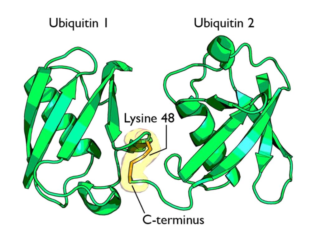
Fig 1 A cartoon representation of a lysine 48-linked diubiquitin molecule. The two ubiquitin chains are shown as green cartoons with each chain labelled. The components of the linkage are indicated and shown as orange sticks. By Rogerdodd (Own work) [CC BY-SA 3.0 (https://creativecommons.org/licenses/by-sa/3.0)], via Wikimedia Commons

The simplest type of chain is homogeneous (IOWs, ubiquitins are linked always at the same site). But many types of mixed and branched chains can also be found.

Let’s start with the most common situation: a poli-ubiquitination of (at least) 4 ubiqutins, linearly linked at K48. This is the common signal for proteasome degradation.

By the way, the 26S proteasome is another molecular machine of incredible complexity, made of more than 30 different proteins. However, its structure and function are not the object of this OP, and therefore I will not deal with them here.

The ubiquitin code is not completely understood, at present, but a few aspects have been well elucidated. Table 1 sums up the most important and well known modes:

Code

Meaning

Polyubiquitination (4 or more) with links at K48 or at K11 Proteasomal degradation
Monoubiqutination (single or multiple) Protein interactions, membrane trafficking, endocytosis
Polyubiquitination with links at K63 Endocytic trafficking, inflammation, translation, DNA repair.
Polyubiquitination with links at K63 (other links) Autophagic degradation of protein substrates
Polyubiquitination with links at K27, K29, K33 Non proteolytic processes
Rarer chain types (K6, K11) Under investigation


However, this is only a very partial approach. A recent bioinformatics paper:

An Interaction Landscape of Ubiquitin Signaling

(Paywall)

Has attempted for the first time a systematic approach to deciphering the whole code, using synthetic diubiquitins (all 8 possible variants) to identify the different interactors with those signals, and they identified, with two different methodologies,  111 and 53 selective interactors for linear polyUb chains, respectively. 46 of those interactors were identified by both methodologies.

The translation

But what “translates” the complex ubiquitin code, allowing ubiquinated proteins to met the right specific destiny? Again, we can refer to the diubiquitin paper quoted above.

How do cells decode this ubiquitin code into proper cellular responses? Recent studies have indicated that members of a protein family, ubiquitin-binding proteins (UBPs), mediate the recognition of ubiquitinated substrates. UBPs contain at least one of 20 ubiquitin-binding domains (UBDs) functioning as a signal adaptor to transmit the signal from ubiquitinated substrates to downstream effectors

But what are those “interactors” identified by the paper (at least 46 of them)? They are, indeed, complex proteins which recognize specific configurations of the “tag” (the ubiquitin chain), and link the tagged (ubiquinated) protein to other effector proteins which implement its final fate, or anyway contribute in deffrent forms to that final outcome.


The basic control of the procedure: the complexity of the ubiquitination process.

So, we have seen that ubiquitin chains work as tags, and that their coded signals are translated by specific interactors, so that the target protein may be linked to its final destiny, or contribute to the desired outcome. But we must still address one question: how is the ubiquitination of the different target proteins implemented? IOWs, what is the procedure that “writes” the specific codes associated to specific target proteins?

This is indeed the first step in the whole process. But it is also the most complex, and that’s why I have left it for the final part of the discussion.

Indeed, the ubiquitination process needs to realize the following aims:

  1. Identify the specific protein to be ubiquitinated
  2. Recognize the specific context in which that protein needs to be ubiquitinated
  3. Mark the target protein with the correct tag for the required fate or outcome

We have already seen that the ubiquitin system is involved in practically all different cellular paths and activities, and therefore we can expect that the implementation of the above functions must be a very complex thing.

And it is.

Now, we can certainly imagine that there are many different layers of regulation that may contribute to the general control of the procedure, specifically epigenetic levels, which are at present poorly understood. But there is one level that we can more easily explore and understand, and it is , as usual, the functional complexity of the proteins involved.

And, even at a first gross analysis, it is really easy to see that the functional complexity implied by this process is mind blowing.

Why? It is more than enough to consider the huge number of different proteins involved. Let’s see.

The ubiquitination process is well studied. It can be divided into three phases, each of which is implemented by a different kind of protein. The three steps, and the three kinds of proteins that implement them, take the name of E1, E2 and E3.
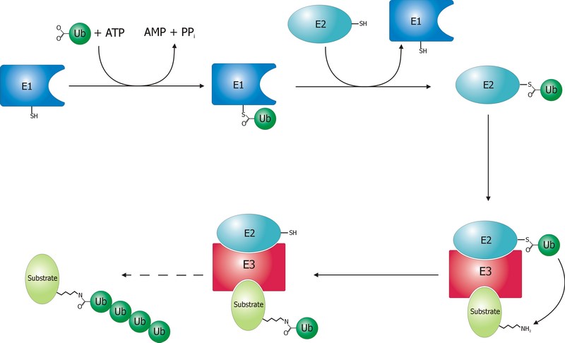
 

Fig. 2 Schematic diagram of the ubiquitylation system. Created by Roger B. Dodd: Rogerdodd at the English language Wikipedia [GFDL (http://www.gnu.org/copyleft/fdl.html) or CC-BY-SA-3.0 (http://creativecommons.org/licenses/by-sa/3.0/)], via Wikimedia Commons

 The E1 step of ubiquitination.

This is the first thing that happens, and it is also the simplest.

E1 is the process of activation of ubiquitin, and the E1 proteins is called E1 ubiquitin-activating enzyme. To put it simply, this enzyme “activates” the ubiquitin molecule in an ATP dependent process, preparing it for the following phases and attaching it to its active site cysteine residue. It is not really so simple, but for our purposes that can be enough.

This is a rather straightforward enzymatic reaction. In humans there are essentially two forms of E1 enzymes, UBA1 and UBA6, each of them about 1000 AAs long, and partially related at sequence level (42%).


The E2 step of ubiquitination.

The second step is ubiquitin conjugation. The activated ubiquitin is transferred from the E1 enzyme to the ubiquitin-conjugating enzyme, or E2 enzyme, where it is attached to a cysteine residue.

This apparently simple “transfer” is indeed a complex intermediate phase. Humans have about 40 different E2 molecules. The following paper:

E2 enzymes: more than just middle men

details some of the functional complexity existing at this level.

Abstract:

Ubiquitin-conjugating enzymes (E2s) are the central players in the trio of enzymes responsible for the attachment of ubiquitin (Ub) to cellular proteins. Humans have ∼40 E2s that are involved in the transfer of Ub or Ub-like (Ubl) proteins (e.g., SUMO and NEDD8). Although the majority of E2s are only twice the size of Ub, this remarkable family of enzymes performs a variety of functional roles. In this review, we summarize common functional and structural features that define unifying themes among E2s and highlight emerging concepts in the mechanism and regulation of E2s.

However, I will not go into details about these aspects, because we have better things to do: we still have to discuss the E3 phase!


The E3 step of ubiquitination.

This is the last phase of ubiquitination, where the ubiquitin tag is finally transferred to the target protein, as initial mono-ubiquitination, or to build an ubiquitin chain by following ubiqutination events. The proteins which implement this final passage are call E3 ubiquitin ligases. Here is the definition from Wikipedia:

A ubiquitin ligase (also called an E3 ubiquitin ligase) is a protein that recruits an E2 ubiquitin-conjugating enzyme that has been loaded with ubiquitin, recognizes a protein substrate, and assists or directly catalyzes the transfer of ubiquitin from the E2 to the protein substrate.

It is rather obvious that the role of the E3 protein is very important and delicate. Indeed it:

  1. Recognizes and links the E2-ubiquitin complex
  2. Recognizes and links some specific target protein
  3. Builds the appropriate tag for that protein (Monoubiquitination, mulptiple monoubiquitination, or poliubiquitination with the appropriate type of ubiquitin chain).
  4. And it does all those things at the right moment, in the right context, and for the right protein.

IOWs, the E3 protein writes the coded tag. It is, by all means, the central actor in our complex story.

So, here comes the really important point: how many different E3 ubiquitin ligases do we find in eukaryotic organisms? And the simple answer is: quite a lot!

Humans are supposed to have more than 600 different E3 ubiquitin ligases!

So, the human machinery for ubiquitination is about:

2 E1 proteins  –  40 E2 proteins – >600 E3 proteins

A real cascade of complexity!

OK, but even if we look at single celled eukaryotes we can already find an amazing level of complexity. In yeast, for example, we have:

1 or 2 E1 proteins  –  11 E2 proteins – 60-100 E3 proteins

See here:

The Ubiquitin–Proteasome System of Saccharomyces cerevisiae

Now, a very important point. Those 600+ E3 proteins that we find in humans are really different proteins. Of course, they have something in common: a specific domain.

From that point of view, they can be roughly classified in three groups according to the specific E3 domain:

  1. RING group: the RING finger domain ((Really Interesting New Gene) is a short domain of zinc finger type, usually 40 to 60 amino acids. This is the biggest group of E3s (about 600)
  2. HECT domain (homologous to the E6AP carboxyl terminus): this is a bigger domain (about 350 AAs). Located at the C terminus of the protein. It has a specific ligase activity, different from the RING   In humans we have approximately 30 proteins of this type.
  3. RBR domain (ring between ring fingers): this is a common domain (about 150 AAs) where two RING fingers are separated by a region called IBR, a cysteine-rich zinc finger. Only a subset of these proteins are E3 ligases, in humans we have about 12 of them.

See also here.

OK, so these proteins have one of these three domains in common, usually the RING domain. The function of the domain is specifically to interact with the E2-ubiquitin complex to implement the ligase activity. But the domain is only a part of the molecule, indeed a small part of it. E3 ligases are usually big proteins (hundreds, and up to thousands of AAs). Each of these proteins has a very specific non domain sequence, which is probably responsible for the most important part of the function: the recognition of the specific proteins that each E3 ligase processes.

This is a huge complexity, in terms of functional information at sequence level.

Our map of the ubiquinating system in humans could now be summarized as follows:

2 E1 proteins  –  40 E2 proteins – 600+ E3 proteins + thousands of specific substrates

IOWs, each of hundreds of different complex proteins recognizes its specific substrates, and marks them with a shared symbolic code based on uniquitin and its many possible chains. And the result of that process is that proteins are destined to degradation by the proteasome or other mechanisms, and that protein interactions and protein signaling are regulated and made possible, and that practically all cellular functions are allowed to flow correctly and smoothly.

Finally, here are two further compoments of the ubuquitination system, which I will barely mention, to avoid making this OP too long.

Ubiquitin like proteins (Ubl):

A number of ubiquitin like proteins add to the complexity of the system. Here is the abstract from a review:

The eukaryotic ubiquitin family encompasses nearly 20 proteins that are involved in the posttranslational modification of various macromolecules. The ubiquitin-like proteins (UBLs) that are part of this family adopt the β-grasp fold that is characteristic of its founding member ubiquitin (Ub). Although structurally related, UBLs regulate a strikingly diverse set of cellular processes, including nuclear transport, proteolysis, translation, autophagy, and antiviral pathways. New UBL substrates continue to be identified and further expand the functional diversity of UBL pathways in cellular homeostasis and physiology. Here, we review recent findings on such novel substrates, mechanisms, and functions of UBLs.

These proteins include SUMO, Nedd8, ISB15, and many others.

Deubiquitinating enzymes (DUBs):

The process of ubiquitination, complex as it already is, is additionally regulated by these enzymes which can cleave ubiquitin from proteins and other molecules. Doing so, they can reverse the effects of ubiquitination, creating a delicately balanced regulatory network. In humans there are nearly 100 DUB genes, which can be classified into two main classes: cysteine proteases and metalloproteases.


By the way, here is a beautiful animation of the basic working of the ubiquitin-proteasome system in degrading damaged proteins:


A summary:

So, let’s try a final graphic summary of the whole ubiquitin system in humans:
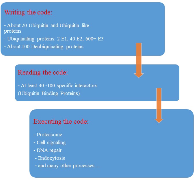
Fig 3 A graphic summary of the Ubiquitin System


Evolution of the Ubiquitin system?

The Ubiqutin system is essentially an eukaryotic tool. Of course, distant precursors for some of the main components have been “found” in prokaryotes. Here is the abstract from a paper that sums up what is known about the prokaryotic “origins” of the system:

Structure and evolution of ubiquitin and ubiquitin-related domains.

(Paywall)

Abstract:

Since its discovery over three decades ago, it has become abundantly clear that the ubiquitin (Ub) system is a quintessential feature of all aspects of eukaryotic biology. At the heart of the system lies the conjugation and deconjugation of Ub and Ub-like (Ubls) proteins to proteins or lipids drastically altering the biochemistry of the targeted molecules. In particular, it represents the primary mechanism by which protein stability is regulated in eukaryotes. Ub/Ubls are typified by the β-grasp fold (β-GF) that has additionally been recruited for a strikingly diverse range of biochemical functions. These include catalytic roles (e.g., NUDIX phosphohydrolases), scaffolding of iron-sulfur clusters, binding of RNA and other biomolecules such as co-factors, sulfur transfer in biosynthesis of diverse metabolites, and as mediators of key protein-protein interactions in practically every conceivable cellular context. In this chapter, we present a synthetic overview of the structure, evolution, and natural classification of Ub, Ubls, and other members of the β-GF. The β-GF appears to have differentiated into at least seven clades by the time of the last universal common ancestor of all extant organisms, encompassing much of the structural diversity observed in extant versions. The β-GF appears to have first emerged in the context of translation-related RNA-interactions and subsequently exploded to occupy various functional niches. Most biochemical diversification of the fold occurred in prokaryotes, with the eukaryotic phase of its evolution mainly marked by the expansion of the Ubl clade of the β-GF. Consequently, at least 70 distinct Ubl families are distributed across eukaryotes, of which nearly 20 families were already present in the eukaryotic common ancestor. These included multiple protein and one lipid conjugated forms and versions that functions as adapter domains in multimodule polypeptides. The early diversification of the Ubl families in eukaryotes played a major role in the emergence of characteristic eukaryotic cellular substructures and systems pertaining to nucleo-cytoplasmic compartmentalization, vesicular trafficking, lysosomal targeting, protein processing in the endoplasmic reticulum, and chromatin dynamics. Recent results from comparative genomics indicate that precursors of the eukaryotic Ub-system were already present in prokaryotes. The most basic versions are those combining an Ubl and an E1-like enzyme involved in metabolic pathways related to metallopterin, thiamine, cysteine, siderophore and perhaps modified base biosynthesis. Some of these versions also appear to have given rise to simple protein-tagging systems such as Sampylation in archaea and Urmylation in eukaryotes. However, other prokaryotic systems with Ubls of the YukD and other families, including one very close to Ub itself, developed additional elements that more closely resemble the eukaryotic state in possessing an E2, a RING-type E3, or both of these components. Additionally, prokaryotes have evolved conjugation systems that are independent of Ub ligases, such as the Pup system.


As usual, we are dealing here with distant similarities, but there is no doubt that the ubiquitin system as we know it appears in eukaryotes.

But what about its evolutionary history in eukaryotes?

We have already mentioned the extremely high conservation of ubiquitin itself.

UBA1, the main E1 enzyme, is rather well conserved from fungi to humans: 60% identity, 1282 bits, 1.21 bits per aminoacid (baa).

E2s are small enzymes, extremely conserved from fungi to humans: 86% identity, for example, for UB2D2, a 147 AAs molecule.

E3s, of course, are the most interesting issue. This big family of proteins behaves in different ways, consistently with its highly specific functions.

It is difficult to build a complete list of E3 proteins. I have downloaded from Uniprot a list of reviewed human proteins including “E3 ubiquitun ligase” in their name: a total of 223 proteins.

The mean evolutionary behavior of this group in metazoa is rather different from protein to protein. However, as a group these proteins exhibit an information jump in vertebrates which is significantly higher than the jump in all other proteins:
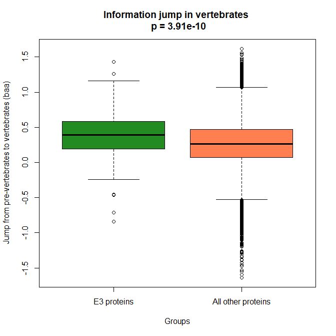
 

Fig. 4 Boxplots of the distribution of human conserved information jump from pre-vertebrates to vertebrates in 223 E3 ligase proteins and in all other human proteins. The difference is highly significant.


As we already know, this is evidence that this class of proteins is highly engineered in the transition to vertebrates. That is consistent with the need to finely regulate many cellular processes, most of which are certainly highly specific for different groups of organisms.

The highest vertebrate jump, in terms of bits per aminoacid, is shown in my group by the E3 ligase TRIM62. also known as DEAR1 (Q9BVG3), a 475 AAs long protein almost absent in pre-vertebrates (best hit 129 bits, 0.27 baa in Branchiostoma belcheri) and which flaunts an amazing jump of 1.433684 baa in cartilaginous fish (810 bits, 1.705263 baa).

But what is this protein? It is a master regulator tumor suppressor gene, implied in immunity, inflammation, tumor genesis.

See here:

TRIM Protein-Mediated Regulation of Inflammatory and Innate Immune Signaling and Its Association with Antiretroviral Activity

and here:

DEAR1 is a Chromosome 1p35 Tumor Suppressor and Master Regulator of TGFβ-Driven Epithelial-Mesenchymal Transition

This is just to show what a single E3 ligase can be involved in!

An opposite example, from the point of view of evolutionary history, is SIAH1, an E3 ligase implied in proteosomal degradation of proteins. It is a 282 AAs long protein, which already exhibits 1.787234 baa (504 bits) of homology in deuterostomes, indeed already 1.719858 baa in cnidaria. However, in fungi the best hit is only 50.8 bits (0.18 baa). So, this is a protein whose engineering takes place at the start of metazoa, and which exhibits only a minor further jump in vertebrates (0.29 baa), which brings the protein practically to its human form already in cartilaginous fish (280 identities out of 282, 99%). Practically a record.

So, we can see that E3 ligases are a good example of a class of proteins which perform different specific functions, and therefore exhibit different evolutionary histories: some, like TRIM62, are vertebrate quasi-novelties, others, like SIAH1, are metazoan quasi-novelties. And, of course, there are other behaviours, like for example BRCA1, Breast cancer type 1 susceptibility protein, a protein 1863 AAs long which only in mammals acquires part of its final sequence configuration in humans.

The following figure shows the evolutionary history of the three proteins mentioned above.
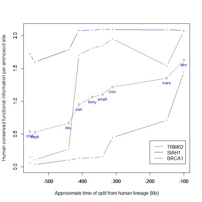
 

Fig. 5 Evolutionary history in metazoa of three E3 ligases (human conserved functional information)


An interesting example: NF-kB signaling

I will discuss briefly an example of how the Ubiquitin system interacts with some specific and complex final effector system. One of the best models for that is the NF-kB signaling.

NK-kB is a transcription factor family that is the final effector of a complex signaling pathway. I will rely mainly on the following recent free paper:

The Ubiquitination of NF-κB Subunits in the Control of Transcription

Here is the abstract:

Nuclear factor (NF)-κB has evolved as a latent, inducible family of transcription factors fundamental in the control of the inflammatory response. The transcription of hundreds of genes involved in inflammation and immune homeostasis require NF-κB, necessitating the need for its strict control. The inducible ubiquitination and proteasomal degradation of the cytoplasmic inhibitor of κB (IκB) proteins promotes the nuclear translocation and transcriptional activity of NF-κB. More recently, an additional role for ubiquitination in the regulation of NF-κB activity has been identified. In this case, the ubiquitination and degradation of the NF-κB subunits themselves plays a critical role in the termination of NF-κB activity and the associated transcriptional response. While there is still much to discover, a number of NF-κB ubiquitin ligases and deubiquitinases have now been identified which coordinate to regulate the NF-κB transcriptional response. This review will focus the regulation of NF-κB subunits by ubiquitination, the key regulatory components and their impact on NF-κB directed transcription.


The following figure sums up the main features of the canonical activation pathway:
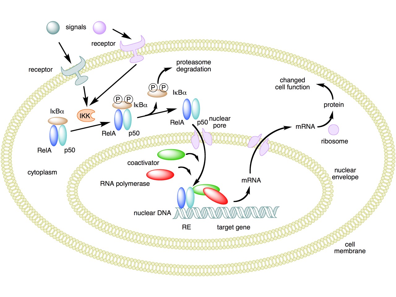
 

Fig. 6 A simple summary of the main steps in the canonical activayion pathway of NF-kB


Here the NF-κB TF is essentially the heterodimer RelA – p50. Before activation, the NF-κB (RelA – p50) dimer is kept in an inactive state and remains in the cytoplasm because it is linked to the IkB alpha protein, an inhibitor of its function.

Activation is mediated by a signal-receptor interaction, which starts the whole pathway. A lot of different signals can do that, adding to the complexity, but we will not discuss this part here.

As a consequence of receptor activation, another protein complex, IκB kinase (IKK), accomplishes the Phosphorylation of IκBα at serines 32 and 36. This is the signal for the ubiquitination of the IkB alpha inhibitor.

This ubiqutination targets IkB alpha for proteosomal degradation. But how is it achieved?

Well, things are not so simple. A whole protein complex is necessary, a complex which implements many different ubiquitinations in different contexts, including this one.

The complex is made by 3 basic proteins:

  • Cul1 (a scaffold protein, 776 AAs)
  • SKP1 (an adaptor protein, 163 AAs)
  • Rbx1 (a RING finger protein with E3 ligase activity, 108 AAs)

Plus:

  • An F-box protein (FBP) which changes in the different context, and confers specificity.

In our context, the F box protein is called beta TRC (605 AAs).
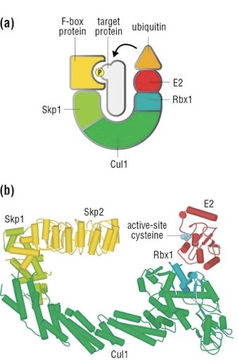
 

Fig. 7 A simple diagram of the SKP1 – beta TRC complex


Once the IkB alpha inhibitor is ubiquinated and degraded in the proteasome, the NF-κB dimer is free to translocate to the nucleus, and implement its function as a transcription factor (which is another complex issue, that we will not discuss).

OK, this is only the canonical activation of the pathway.

In the non canonical pathway (not shown in the figure) a different set of signals, receptors and activators acts on a different NF-κB dimer (RelB – p100). This dimer is not linked to any inhibitor, but is itself inactive in the cytoplasm. As a result of the signal, p100 is phosphorylated at serines 866 and 870. Again, this is the signal for ubiquitination.

This ubiquitination is performed by the same complex described above, but the result is different. P100 is only partially degraded in the proteasome, and is transformed into a smaller protein, p52, which remains linked to RelB. The RelB – p52 dimer is now an active NF-κB Transcription Factor, and it can relocate to the nucleus and act there.

But that’s not all.

  • You may remember that RelA (also called p 65) is one of the two components of NF-kB TF in the canonical pathway (the other being p 50). Well, RelA is heavily controlled by ubiquitination after it binds DNA in the nucleus to implement its TF activity. Ubiquitination (a very complex form of it) helps detachment of the TF from DNA, and its controlled degradation, avoiding sustained expression of NF-κB-dependent genes. For more details, see section 4 in the above quoted paper: “Ubiquitination of NF-κB”.
  • The activation of IKK in both the canonical and non canonical pathway after signal – receptor interaction is not so simple as depicted in Fig. 6. For more details, look at Fig. 1 in this paper: Ubiquitin Signaling in the NF-κB Pathway. You can see that, in the canonical pathway, the activation of IKK is mediated by many proteins, including TRAF2, TRAF6, TAK1, NEMO.
  • TRAF2 is a key regulator on many signaling pathways, including NF-kB. It is an E3 ubiquitin ligase. From Uniprot:  “Has E3 ubiquitin-protein ligase activity and promotes ‘Lys-63’-linked ubiquitination of target proteins, such as BIRC3, RIPK1 and TICAM1. Is an essential constituent of several E3 ubiquitin-protein ligase complexes, where it promotes the ubiquitination of target proteins by bringing them into contact with other E3 ubiquitin ligases.”
  • The same is true of TRAF6.
  • NEMO (NF-kappa-B essential modulator ) is also a key regulator. It is not an ubiquinating enzyme, but it is rather heavily regulated by ubiquitination. From Uniprot: “Regulatory subunit of the IKK core complex which phosphorylates inhibitors of NF-kappa-B thus leading to the dissociation of the inhibitor/NF-kappa-B complex and ultimately the degradation of the inhibitor. Its binding to scaffolding polyubiquitin seems to play a role in IKK activation by multiple signaling receptor pathways. However, the specific type of polyubiquitin recognized upon cell stimulation (either ‘Lys-63’-linked or linear polyubiquitin) and its functional importance is reported conflictingly.”
  • In the non canonical pathway, the activation of IKK alpha after signal – receptor interaction is mediated by other proteins, in particular one protein called NIK (see again Fig. 1 quoted above). Well, NIK is regulated by two different types of E3 ligases, with two different types of polyubiquitination:
    • cIAP E3 ligase inactivates it by constant degradation using a K48 chain
    • ZFP91 E3 ligase stabilizes it using a K63 chain

See here:

Non-canonical NF-κB signaling pathway.

In particular, Fig. 3

These are only some of the ways the ubiquitin system interacts with the very complex NF-kB signaling system. I hope that’s enough to show how two completely different and complex biological systems manage to cooperate by intricate multiple connections, and how the ubiquitin system can intervene at all levels of another process. What is true for the NF-kB signaling pathway is equally true for a lot of other biological systems, indeed for almost all basic cellular processes.

But this OP is already too long, and I have to stop here.

As usual, I want to close with a brief summary of the main points:

  1. The Ubiquitin system is a very important regulation network that shows two different signatures of design: amazing complexity and an articulated semiotic structure.
  2. The complexity is obvious at all levels of the network, but is especially amazing at the level of the hundreds of E3 ligases, that can recognize thousands of different substrates in different contexts.
  3. The semiosis is obvious in the Ubiquitin Code, a symbolic code of different ubiquitin configurations which serve as specific “tags” that point to different outcomes.
  4. The code is universally implemented and shared in eukaryotes, and allows control on almost all most important cellular processes.
  5. The code is written by the hundreds of E3 ligases. It is read by the many interactors with ubiquitin-binding domains (UBDs).
  6. The final outcome is of different types, including degradation, endocytosis, protein signaling, and so on.
  7. The interaction of the Ubiquitin System with other complex cellular pathways, like signaling pathways, is extremely complex and various, and happens at many different levels and by many different interacting proteins for each single pathway.

PS:

Thanks to DATCG for pointing to this video in three parts by Dr. Raymond Deshaies, was Professor of Biology at the California Institute of Technology and an Investigator of the Howard Hughes Medical Institute. On iBiology Youtube page:

A primer on the ubiquitin-proteasome system


Cullin-RING ubiquitin ligases: structure, structure, mechanism, and regulation


Targeting the ubiquitin-proteasome system in cancer

Comments
#410 and #409 Gpuccio, Yes! It's fascinating "stuff" our neural capacity, plasticity and change factors. Including all forms of stress, embryonic development and formation. Long-term memory, epigenetic regulatory systems and well, our little ubiquitin friends. "I think that this field is in great expansion, and maybe we can see something more specific in a short time." I agree! Very interested in this area. Hope to devote more time to study these areas in future. I'm encouraged by the advancements in research being made at a rapid pace.DATCG
March 16, 2018
March
03
Mar
16
16
2018
11:20 PM
11
11
20
PM
PST
#411 Gpuccio, re: CK1alpha and Slimb Thanks! It seems UB E3 Slimb being a little "slower" makes sense due to species specific needs? The pesky human brain for example? On Condensins, Chromosomes, and beautiful DNA packaging and compression... "Let's put it among our future plans!" Ahaha! :) Your list is growing Gpuccio!DATCG
March 16, 2018
March
03
Mar
16
16
2018
11:06 PM
11
11
06
PM
PST
DATCG at #399: "Q: if CK1alpha is highly conserved, then is UBLigase-E3 Slimb highly conserved with it?" Yes they are both highly conserved in metazoa. CK1alpha (337 AAs): the human protein shows 71% identities and 84% positives in Fungi (483 bits, 1.433 baa), and reaches a practically complete homology (99% identities, 100% positives) in Cartilaginous Fish. Amazing! Slimb (542 AAs) is just a little "slower": 40% idenitites and 62% positives in Fungi, 91% and 93% in Cartilaginous Fish. Fascinating data about the condensin complexes. This issue of chromosome and chromatin structure certainly deserves some in-depth analysis. Let's put it among our future plans! :)gpuccio
March 16, 2018
March
03
Mar
16
16
2018
04:49 PM
4
04
49
PM
PST
DATCG at #393: Nice stuff about ubiquitin and addiction. By the way, the linked Table 1 is really amazing! :)gpuccio
March 16, 2018
March
03
Mar
16
16
2018
04:24 PM
4
04
24
PM
PST
DATCG at #393: Fascinating facts about long term memory. I think that this field is in great expansion, and maybe we can see something more specific in a short time. The working of the brain and nervous system is probably to be explained, as far as that is possible, at two different levels: a) The network of connections between neurons (and other cell types). This is amazing, if we think that we have about 10^11 neurons, and maybe 10^15 neuron connections. Those are big numbers, indeed. Any expert in hardware and software engineering knows all to well how important it is to have the right connections. And neuronal connections are dynamic, they can change and be rewired. b) But even more amazing is the biochemical plasticity of all that happens in neurons, and especially in synapses. And out friend ubiquitin, as you have shown, is critically linked to all this. I think that the only luck for neo-darwinists as far as the central nervous system is concerned is that we really understand too litlle of how it works. At least for the moment.gpuccio
March 16, 2018
March
03
Mar
16
16
2018
04:20 PM
4
04
20
PM
PST
Upright BiPed: All of you, DATCG, Dionisio and you, have given great contributions! :) I was just focusing on DATG because his comments have become really prominent in the last phase of the discussion...gpuccio
March 16, 2018
March
03
Mar
16
16
2018
11:11 AM
11
11
11
AM
PST
GP at 404 Agreed -- and Dio's contributions as well. I don't know about this DATCG character. We'll have to keep our eyes on him/her. He/she appears to be exceptionally bright. Clearly not his/her first rodeo in this area. :) :) :)Upright BiPed
March 16, 2018
March
03
Mar
16
16
2018
09:19 AM
9
09
19
AM
PST
#404 Gpuccio, and Dionisio contributions add even more! I've not come close to reviewing all of his papers! Simply not enough time. It's a cornucopia of Ubiquitin fruit ;-)DATCG
March 16, 2018
March
03
Mar
16
16
2018
08:02 AM
8
08
02
AM
PST
#401-403 Gpuccio, Thanks, that illuminates the field of Epigenetics. I'd not considered Transcription's dependence on Epigenetic factors. Then there's so many other networks dependent upon Epigenetics. The 75% threshold by Dan Graur was precarious from the start. But I think he built that artificial wall based upon what he must have for neo-Darwinian faith to continue. By creating this in his anger and stubborn attitude he's erected what appears to be a Humpty Dumpty Wall made of cards. and... “If Evolution is wrong, ENCODE is right” ah :) Now you have juxtaposed a good turnabout is fair play. Dan and his mirror... as ENCODE proceeds and non-coded regions are explored with new functions found every day around the world. Mirror, mirror on the wall, who is right after all? Is it Darwin, is it Dan? Do unguided lots make up plans? Mirror, mirror on the wall, is neo-Darwinism due to fall? Humpty Dumpty Darwin's game, does blind search fall in shame?DATCG
March 16, 2018
March
03
Mar
16
16
2018
07:54 AM
7
07
54
AM
PST
Upright BiPed at # 397: "I’m trying to catch up, but it seems almost impossible." I can understand you. Sometimes it seems almost impossible to me to catch up with myself! :) However, it seems that adding DATCG to myself works combinatorially in fully unexpected ways. The results are really scary! :)gpuccio
March 16, 2018
March
03
Mar
16
16
2018
06:54 AM
6
06
54
AM
PST
DATCG at #392: “If ENCODE is right, Evolution is wrong” What a pity! If it had been the other way round: "If Evolution is wrong, ENCODE is right" we could be certain that ENCODE is right! :) However, I agree with you that maybe some non coding DNA could be non functional, but it's certainly not 75%! This 4D Nucleome Project is extremely interesting. I am sure that the 3D dynamic structure of chromatin and its constant modifications in time are one of the most important keys to understand something which goes beyond a mere accumulation od fetails. Hi-C (and its variants) is a really promising technique. The real primary aim is to understand how TFs work, their combinatorial nature, their ability to form chromatin loops and to connect distant parts of the genome in functional complexes. At that level, we are really just beginning to understand things.gpuccio
March 16, 2018
March
03
Mar
16
16
2018
06:50 AM
6
06
50
AM
PST
DATCG at #391: "Does it logically follow that Ubiquitin is fully dependent upon Epigenetic layers of meta-code to function in all these different areas covered so far in this one OP?" Well, probably almost everything is under the control of epigenetic layers, because all transcription is fully dependent upon them. But ubuquitin has a definite role on epigenetic layers, as shown for example by its many roles with histones. It's not a cse that we find ever more often the term "cross-talk" in biological papers. One thing is astonishingly clear: the cell has many, many independent layers of regulations, and all of them are constantly influencing one another and exchanging information. I think this is unprecedented, even in human programming and engineering.gpuccio
March 16, 2018
March
03
Mar
16
16
2018
06:16 AM
6
06
16
AM
PST
Especially if that network works by coded symbols, like the different types of signals implemented by ubiquitin chains. Especially if the network is made by hundreds and hundreds of specific sub-networks. Especially if the network controls not one, but tons of different complex functions, practically every function we can imagine.
Terrifying -- if it’s your job to make sure there are never enough dissenters, that they might change the paradigm. :) Going to need some extra dogma. I suspect some shame, threats, and group enforcement will come in handy as well.Upright BiPed
March 15, 2018
March
03
Mar
15
15
2018
11:04 PM
11
11
04
PM
PST
Packing a Genome, Step-by-Step - Condensin II Just to cool. While the video or "steps" do not say it. Somewhere, where there's regulation, there's Ubiquitination. Remember Condensin II and E3-ligase Slimb regulation in coordination with phosphorlation. - Ooops @399, sorry - missed a closing Bold Font highlight.DATCG
March 15, 2018
March
03
Mar
15
15
2018
09:40 PM
9
09
40
PM
PST
Walking thru time on the 4D Nucleome Project, I ventured out a bit to see if I could find Ubiquitin involvement in different areas. Here's an interesting related area to review... Drosophila Casein Kinase I Alpha Regulates Homolog Pairing and Genome Organization by Modulating Condensin II Subunit Cap-H2 Levels PLoS Genet. 2015 Feb;11(2): e1005014.Published online 2015 Feb 27. doi: 10.1371/journal.pgen.1005014 Huy Q. Nguyen, Jonathan Nye, Daniel W. Buster, Joseph E. Klebba, Gregory C. Rogers,and Giovanni Bosco, R. Scott Hawley, Editor Abstract
The spatial organization of chromosomes within interphase nuclei is important for gene expression and epigenetic inheritance. Although the extent of physical interaction between chromosomes and their degree of compaction varies during development and between different cell-types, it is unclear how regulation of chromosome interactions and compaction relate to spatial organization of genomes. Drosophila is an excellent model system for studying chromosomal interactions including homolog pairing. Recent work has shown that condensin II governs both interphase chromosome compaction and homolog pairing and condensin II activity is controlled by the turnover of its regulatory subunit Cap-H2. Specifically, Cap-H2 is a target of the SCFSlimb E3 ubiquitin-ligase which down-regulates Cap-H2 in order to maintain homologous chromosome pairing, chromosome length and proper nuclear organization. Here, we identify Casein Kinase I alpha(CK1-alpha) as an additional negative-regulator of Cap-H2. CK1alpha-depletion stabilizes Cap-H2 protein and results in an accumulation of Cap-H2 on chromosomes. Similar to Slimb mutation, CK1alpha depletion in cultured cells, larval salivary gland, and nurse cells results in several condensin II-dependent phenotypes including dispersal of centromeres, interphase chromosome compaction, and chromosome unpairing. Moreover, CK1alpha loss-of-function mutations dominantly suppress condensin II mutant phenotypes in vivo. Thus, CK1alpha facilitates Cap-H2 destruction and modulates nuclear organization by attenuating chromatin localized Cap-H2 protein.
Introduction
Interphase genome organization in eukaryotic cells is non-random [1,2,3]. Indeed, organization of the genome is crucial because it influences nuclear shape and processes such as DNA repair and replication, as well as gene expression [4, 5, 6]. While chromosomes are highly organized within the nucleus, they must also remain extremely dynamic. Chromosome dynamics facilitate events that occur not only during cell division, but also during interphase, when cells respond to developmental and environmental cues that require changes in gene expression. Interphase events include trans-interactions such as homolog pairing, chromosome remodeling and compaction, and DNA looping. Although numerous studies using Fluorescent In-Situ Hybridization (FISH), live cell imaging, and chromosome conformation capture techniques have revealed the three-dimensional (3D) organization of genomes, much remains to be discovered regarding the factors that govern the overall conformation of interphase chromosomes. An equally important task is to identify the molecular mechanisms that regulate and maintain specific 3D genome organizational states. Condensin complexes are highly conserved from bacteria to humans [7,8,9] and have been identified as key drivers of genome organization [10]. Eukaryotes have two condensin complexes, condensin I and II, which share the core SMC2 and SMC4 (Structural Maintenance of Chromosomes) subunits but differ in their non-SMC Chromosome Associated Protein (CAP) subunits. Condensins have long been known to play vital roles in shaping mitotic chromosomes. While condensin I promotes lateral chromosome compaction, condensin II promotes axial compaction; both of which are necessary for faithful mitotic condensation and chromosome segregation [11]. Condensins also display different localization patterns: condensin I only associates with mitotic chromosomes, whereas condensin II is present in the nucleus, where it is bound to chromatin throughout the cell cycle [12,13,14,15]
Fascinating. So where Ubiquitin? Here, Slimb E3 ligase... Ubiquitin E3 coordination with Phosphorlation and Degradation
note: ? = alpha subunit Moreover, Cap-H2 protein levels are controlled by the SCFSlimb ubiquitin-ligase, maintaining low levels of Cap-H2 in vivo and in cultured Drosophila cells [20]. Interestingly, Slimb(E3) recognizes its target proteins through a phosphodegron motif [29], suggesting that one or more kinases must phosphorylate Cap-H2 before Slimb can target it for destruction. A Slimb-binding site consensus sequence (DSGXXS) exists in the extreme C-terminus of Cap-H2 and deletion of this region renders Cap-H2 non-degradable [20]. As expected for a Slimb substrate, Cap-H2 protein mobility on SDS-PAGE was sensitive to phosphatase treatment, suggesting that Cap-H2 is phosphorylated [20]. Given that Cap-H2 protein levels may be regulated by its phosphorylation state, we set out to identify kinases that target Cap-H2 for Slimb recognition and that lead to its degradation. We show that in Drosophila cultured S2 cells, Casein Kinase I alpha (CK1?) depletion results in the hypercondensation of interphase chromatin in a condensin II-dependent manner. We also found that CK1? and condensin II genetically interact in vivo, and that CK1? depletion leads to Cap-H2 protein enrichment on polytene and cultured cell chromosomes. Similar to Slimb(E3) depletion [20], CK1? depletion also results in stabilization of Cap-H2 protein in cultured cells. Our findings further elucidate the mechanism by which Cap-H2, and thus condensin II, is regulated and contribute significantly to our understanding of how interphase genome organization, homolog pairing, and chromosome compaction is modulated.
Results Casein Kinase I alpha is required for interphase chromatin reorganization
Previously, we discovered that the Cap-H2 subunit of condensin II is a SCFSlimb ubiquitination-target in Drosophila cells [20]. In a whole genome RNAi screen, Slimb was also identified as a homolog pairing-promoting factor, and it was shown to affect pairing in a Cap-H2 dependent manner[18]. In cultured S2 and Kc cells, depletion of SCFSlimb components Slimb, Cul-1 and SkpA prevents Cap-H2 degradation and leads to condensin II hyperactivation during interphase and the remodeling of each chromosome into a compact globular structure (Fig. 1A-C). Based on their overall appearance, we refer to these hypercondensed chromosomes as “chromatin-gumballs” (Fig. 1A). Overexpression of a GFP tagged wild type Cap-H2 also induces this phenotype [20]. Since phosphorylation of the Slimb-binding domain within its substrates is required for Slimb binding [29], we reasoned that depletion of a kinase involved in this pathway would also stabilize Cap-H2 and phenocopy the effect on chromatin remodeling observed after Slimb depletion.
CK1alpha Highly Conserved Kinase CK1?=CK1alpha
CK1? is a highly conserved serine/threonine kinase involved in Wnt signaling pathways, DNA repair, cell cycle progression, and mRNA metabolism [35,47,48]. Identification of CK1? furthers our understanding of the mechanisms by which condensin II is regulated. The chromodomain protein Mrg15 is involved in the loading of Cap-H2, while the E3 Ubiquitin ligase, SCFSlimb ubiquitylates Cap-H2, removing it from chromatin and targeting it for proteasomal degradation [20,21]. Phosphorylation is known to be a prerequisite for Slimb recognition of its target proteins
Q: if CK1alpha is highly conserved, then is UBLigase-E3 Slimb highly conserved with it?
It is tempting to speculate that cytokine signaling could trigger the activation of a condensin II antagonist, leading to the decrease in condensin II activity. This would lead to decondensation of chromatin allowing STAT5 access to DNA. Our findings in the Drosophila model suggest that similar interphase condensin II functions may be at play, and CK1? along with Slimb are critical regulators of this condensin II activity. However, at present it is not known if mammalian condensin II activity is regulated by Slimb or CK1?, and it should be noted that mouse and human Cap-H2 do not have clear Slimb binding consensus sequences. It will be of great value to identify additional kinases that may collaborate with CK1? and Slimb to negatively regulate Drosophila condensin II activity, and to further elucidate the biological significance of this interphase condensin II function in Drosophila and other species.
OK, so Condensin II, next up a cool video. Packaging Pathways. .DATCG
March 15, 2018
March
03
Mar
15
15
2018
09:06 PM
9
09
06
PM
PST
Gpuccio @395 "So, how is it that no one from the other field has writeen one sinlge word here to try to explain how random variation and natural selection can do this?" Good question, where are the neo-Darwinist? I suspect they stay away for purposes of not making your excellent OPs legitimate - as in recognized not only by themselves, but in eyes of their own followers. Think of it. If they actually engage you - they can lose. And their followers might see your logic as correct. They cannot bear that possible outcome. And I suspect Hunt got a hint to back away. I could be wrong, but am surprised he'd back off for any other reason. Surely he can mount a defense of group II introns and spliceasome evolution, right? By not engaging, they hope Intelligent Design goes away. It's not, it's only growing. And more bright minds are learning every day a new way of seeing life as a result of Design. Discovery Institute Summer Seminars on Intelligent Design July 6-14 .DATCG
March 15, 2018
March
03
Mar
15
15
2018
07:54 PM
7
07
54
PM
PST
Simply outstanding work by the two of you. I'm trying to catch up, but it seems almost impossible. Great job.Upright BiPed
March 15, 2018
March
03
Mar
15
15
2018
05:15 PM
5
05
15
PM
PST
DATCG at #390: "How many future OPs are you entertaining now? " Indeed, I am thinking about 3 or 4 different possibilities. In the end, I will probably follow some sudden "inspiration"! :) The "prokaryote to eukaryote transition" is a fascinating issue. What a pity that we have no precise idea of when it happened, and least of all a reasonable early tree of eukaryotes! I think that both my OP on the spliceosome and this one about ubiquitin are good examples of highly specific eukaryotic machinery. But of course, there are many others! :) "This is where I disagreed with Arthur Hunt and thought BLASTing information was critical in reviewing informational jumps. Why he was critical of it, remains a mystery. Perhaps because it hits close to home." I disagree with Arthur Hunt too, as much as it is possible to disagree with someone who has not really expressed his thoughts. :) BLAST is a wonderful tool for us IDists. Neo-darwinists use it mostly to find vague distant homologies. But we can and do use it to detect functional information, which is much more interesting! OK, it's late now here in Italy. I will come back tomorrow.gpuccio
March 15, 2018
March
03
Mar
15
15
2018
04:53 PM
4
04
53
PM
PST
DATCG at #389: "Random mutations are the enemy, not the blind, unguided builder of such highly integrated, tightly regulated, Functionally Organized & Highly Coordinated Complex Systems." Of course. The idea that a complex regulation network may arise from random mutations and natural selection is ridiculous! Especially if that network works by coded symbols, like the different types of signals implemented by ubiquitin chains. Especially if the network is made by hubdreds and hundreds of specific sub-networks. Especially is the network control not one, but tons of different complex functions, practically every function we can imagine. So, how is it that no one from the other field has writeen one sinlge word here to try to explain how random variation and natural selection can do this? :) "The material covered is an avalanche of specified information, overwhelming any highly trained team of scientist and lab techs to keep up with." You bet! Just the 600+ E3 ligases are an example of thousands and thousands, maybe hundreds of thousands, of functional bits whose function is to recognize all the specific target proteins, thousands of them, and tag them in the correct way in each appropriate condition. Let's remember that about 5% of the whole protein coding genome is implied in the ubuquitin network! "OK, I just stated the obvious. But sometimes the obvious must be stated" Absolutely! :) When no one seems to have the courage to deny the absurd, stating the obvious is probably the only salvation. :)gpuccio
March 15, 2018
March
03
Mar
15
15
2018
04:45 PM
4
04
45
PM
PST
Part of Drug Abuse and Addiction is Conditioning. Habit forming over time. While certainly dealing with different aspects of neural development and different areas related to addiction, I'd now expect to find UB or UPS, and DUBS in some role. So, searched on UB and Addiction. And oddly enough found the Review Article below in the same journal... Roles of the ubiquitin proteasome system in the effects of drugs of abuse Front. Mol. Neurosci., 06 January 2015 | https://doi.org/10.3389/fnmol.2014.00099 Nicolas Massaly, Bernard Francès and Lionel Moulédous What I did not expect immediately to see, but makes absolute sense is the overlapping of Memory in conjunction with addiction and UB.
Because of its ability to regulate the abundance of selected proteins the ubiquitin proteasome system (UPS) plays an important role in neuronal and synaptic plasticity. As a result various stages of learning and memory depend on UPS activity. Drug addiction, another phenomenon that relies on neuroplasticity, shares molecular substrates with memory processes. However, the necessity of proteasome-dependent protein degradation for the development of addiction has been poorly studied. Here we first review evidences from the literature that drugs of abuse regulate the expression and activity of the UPS system in the brain. We then provide a list of proteins which have been shown to be targeted to the proteasome following drug treatment and could thus be involved in neuronal adaptations underlying behaviors associated with drug use and abuse. Finally we describe the few studies that addressed the need for UPS-dependent protein degradation in animal models of addiction-related behaviors.
Interesting, regulation after Drug Exposure(B) in Figure 1 UPS components regulated(see B) after Drug Exposure Figure 1. The Ubiquitin Proteasome System and its components regulated after drug exposure.
(A) Schematic representation of the Ubiquitin Proteasome System. The external and internal rings constitute the 20S proteasome. The lid and base constitute the 19S regulatory complex. In some cases, it can be replaced by the PA28 or 11S regulatory complex, constituted of a single ring of 7 subunits. (B) Classification of the UPS components found to be regulated after drug exposure.
All drugs of abuse can thus affect the expression and abundance of key UPS proteins.
However, the data reported above are only descriptive. Moreover, UPS components are affected differently depending on the drug type, its method of administration, the duration of the treatment and the cell type or brain region considered (Table 1). Complementary studies have also found that drugs of abuse modify the activity of the UPS in parallel with changes in the expression of its various components. Indeed morphine was demonstrated to inhibit the activity of the 20S proteasome in human neuroblastoma cells, with neuroprotective consequences (Rambhia et al., 2005). On the contrary, PKC-dependent inhibition of the UPS was linked to the autophagy-mediated toxicity of methamphetamine in dopaminergic neurons (Lin et al., 2012). In addition it has been proposed that the higher toxicity of methamphetamine compared to cocaine was due to its long inhibitory effect on proteasome activity (Dietrich et al., 2005). Finally, a recent study demonstrated that chronic ethanol induces toxicity in mice through a Toll-like receptor 4-dependent impairment of the UPS (Pla et al., 2014).
Balance between Protein Synthesis and Degradation again
This deleterious effect of UPS blockade on long term changes in neurons has been suggested to be due to an alteration in the balance between protein synthesis and degradation (Fonseca et al., 2006). Indeed the authors showed that the deleterious effects produced by inhibiting either protein synthesis or degradation on LTP can be reversed by inhibition of the two processes at the same time. In addition to synaptic proteins the UPS is also involved in the regulation of the activity of transcription factors, thus revealing a close relationship between protein synthesis and proteasome action. For example I?B and CREM (cAMP-responsive element modulator), repressors of the transcription factors NF-?B and CREB (cAMP response element binding) respectively, can be ubiquitinated and degraded by the UPS (Woo et al., 2010; Liu and Chen, 2011). In that sense the UPS clearly plays a major role in the regulation of protein turnover implicated in neuronal plasticity acting directly through the degradation of some proteins and indirectly through the modulation of transcriptional activity and protein synthesis.
Oh, the authors recognize Jarome, et al., previous work in UPS regulation and memory(LTM), but I'm moving on to another paragraph.
The precise mechanisms underlying the involvement of the proteasome in memory are just beginning to be discovered but it is now clearly established that, in addition to protein synthesis, neuronal protein degradation by the UPS is a mandatory process to create, store and maintain memories and in that sense participates to adaptive behaviors of mammals. Since drug addiction shares common mechanisms with memory processes (Hyman et al., 2006; Milton and Everitt, 2012) it is important to question the role of the UPS in the long term effects of drugs of abuse such as opioids, stimulants, ethanol, nicotine and cannabinoids.
Indeed, very interesting material.
In the case of opioids, it was shown in a cellular model that a prolonged 72 h morphine treatment modifies the abundance of two proteasome subunits (?3 and ?6) (Neasta et al., 2006). In vivo, intra-cerebro-ventricular (icv) infusion of morphine for 72 h results in an increase in the tyrosine-phosphorylated form of the ?4 subunit in the rat frontal cerebral cortex (Kim et al., 2005). A longer intermittent treatment (2 weeks) produces a decrease in the amount of the DUB Ubiquitin C-terminal hydrolase L-1 in the nucleus accumbens (Nacc) (Li et al., 2006). 4 days after morphine withdrawal, the quantity of this enzyme, as well as that of the ?3 subunit of the proteasome, increases in rat dorsal root ganglia (Li et al., 2009). Similarly, chronic treatment (90 days) and drug withdrawal have been shown to have opposite effects on the amount of ?5 subunit in the Nacc of rhesus monkeys (Bu et al., 2012). The levels of Ubiquitin-conjugating enzyme E2 and of Ubiquitin C-terminal hydrolase L-3 are also modulated in this model. Finally, in a morphine-induced conditioned place preference (CPP) paradigm which tests the rewarding properties of the drug, both development, extinction and re-instatement are accompanied by a down-regulation of several DUBs and ? and ? subunits (Lin et al., 2011).
Hmmmm, now, what of legal pharmaceuticals? Not to beat up on Big Pharma, but what of psychotropic medicines intended for good that are suddenly removed from a patient? What happens with build up and changes in different areas? How does the brain and neural network balance after sudden removal, including UPS, DUBS and other regulatory systems involved at cognition, perception and memory? What remains unchanged? What is inherited Epigenetic changes passed down to offspring? Table 1. UPS-related molecular and cellular consequences of the treatment with drugs of abuse. That's a load of UPS regulatory functions and consequences by drugs of abuse. Wonder if similar studies exist for legal drugs showing similar areas of changes and modifications for public access. .DATCG
March 15, 2018
March
03
Mar
15
15
2018
02:26 PM
2
02
26
PM
PST
#387 Gpuccio, back on topic Memory and UPS, a research review by Timothy Jarome in a paper he published 2014. Open Access. Ubiquitin Role in Long Term Memory Formation with Protein Degradation and Synthesis REVIEW ARTICLE Front. Mol. Neurosci., 26 June 2014 | https://doi.org/10.3389/fnmol.2014.00061 Timothy J. Jarome and Fred J. Helmstetter
Long-term memory (LTM) formation requires transient changes in the activity of intracellular signaling cascades that are thought to regulate new gene transcription and de novo protein synthesis in the brain. Consistent with this, protein synthesis inhibitors impair LTM for a variety of behavioral tasks when infused into the brain around the time of training or following memory retrieval, suggesting that protein synthesis is a critical step in LTM storage in the brain. However, evidence suggests that protein degradation mediated by the ubiquitin-proteasome system (UPS) may also be a critical regulator of LTM formation and stability following retrieval. This requirement for increased protein degradation has been shown in the same brain regions in which protein synthesis is required for LTM storage. (balance and trade-off of keeping memory retainable across time) Additionally, increases in the phosphorylation of proteins involved in translational control parallel increases in protein polyubiquitination and the increased demand for protein degradation is regulated by intracellular signaling molecules thought to regulate protein synthesis during LTM formation. (balance must be maintained again) In some cases inhibiting proteasome activity can rescue memory impairments that result from pharmacological blockade of protein synthesis, suggesting that protein degradation may control the requirement for protein synthesis during the memory storage process. Amazing, balance Results such as these suggest that protein degradation and synthesis are both critical for LTM formation and may interact to properly “consolidate” and store memories in the brain. Here, we review the evidence implicating protein synthesis and degradation in LTM storage and highlight the areas of overlap between these two opposing processes. (Opposing Processes = Balancing Act) We also discuss evidence suggesting these two processes may interact to properly form and store memories. LTM storage likely requires a coordinated regulation between protein degradation and synthesis at multiple sites in the mammalian brain.
Amazing stuff, this seesaw of regulation, synthesis and degradation.
Recently, attention has turned to the potential role of protein degradation in learning-dependent synaptic plasticity. Indeed, there is now convincing evidence that UPS-mediated protein degradation is likely involved in various different stages of memory storage. However, while some studies have suggested potential roles for protein degradation in long-term memory (LTM) formation and storage (Kaang and Choi, 2012), one intriguing question is whether protein degradation is linked to the well-known transcriptional and translational alterations thought to be critical for memory storage in the brain (Johansen et al., 2011). Here, we discuss evidence demonstrating a role for protein degradation and synthesis in the long-term storage of memories in the mammalian brain, highlighting instances in which a requirement for protein degradation correlates with a requirement for protein synthesis. Additionally, we discuss evidence suggesting that both protein degradation and synthesis may be regulated by CaMKII signaling during LTM formation. Collectively, we propose that LTM storage requires coordinated changes in protein degradation and synthesis in the brain, which may be primarily controlled through a CaMKII-dependent mechanism.
Had not read down this far but oh so cool... What Comes First, Degradation or Synthesis?
A majority of the studies discussed here reveal a strong correlation between protein degradation and synthesis during LTM formation. This leads to one important question: Which comes first? While the exact relationship between protein degradation and synthesis during memory formation currently remains equivocal, the available evidence suggests that protein degradation likely regulates protein synthesis. For example, fear conditioning leads to an increase in polyubiquitinated proteins being targeted for degradation by the proteasome (Jarome et al., 2011). While a majority of the proteins being targeted by the proteasome for degradation remain unknown, the RNAi-induced Silencing Complex (RISC) factor MOV10 has been identified as a target of the proteasome during increases in activity-dependent protein degradation in vitro (Banerjee et al., 2009) and following behavioral training and retrieval in vivo (Jarome et al., 2011). Increases in the degradation of MOV10 are associated with increased protein synthesis in vitro, suggesting that the proteasome could regulate protein synthesis during LTM formation through the removal of translational repressor proteins such as various RISC factors. However, it is currently unknown if the selective degradation of MOV10, or any RISC factor, is critical for memory formation in neurons. Nonetheless, studies such as these provide indirect evidence that protein degradation by the UPS could regulate protein synthesis during memory formation in the brain.
The balancing act and pre-programmed responses to conditioning possibly exert factors on enhanced protein synthesis processing.
Some of the best evidence that protein degradation may be upstream of protein synthesis during memory storage comes from studies examining memory reconsolidation following retrieval. For example, inhibiting proteasome activity can prevent the memory impairments that normally result from post-retrieval blockade of protein synthesis in the hippocampus, amygdala, and nucleus accumbens (Lee et al., 2008; Jarome et al., 2011; Ren et al., 2013) as well as during LTF in aplysia (Lee et al., 2012), suggesting that protein degradation is upstream of protein synthesis during memory reconsolidation. This remains some of the best evidence directly linking protein degradation to protein synthesis during memory storage, but it is possible that the rescue of memory impairments in the face of protein synthesis inhibition may occur as an indirect consequence of blocking protein degradation rather than a direct effector.
Now, how is ubiquitin code and protocol utilized(or inhibited, modified) in Addictions, Drug Abuse or legal drugs for reducing pain? I'll move this to a new comment.DATCG
March 15, 2018
March
03
Mar
15
15
2018
02:20 PM
2
02
20
PM
PST
Continued from #391 ... offtopic a bit, but looking ahead. On possible links between Ubiqutin and Epigenetic roles - does this lead to decimation of Dan Graur's argument for large amount of "Junk" DNA sill existing? According to his last paper? In his last retort he states at least 75% of our DNA must be JUNK. I'm not entertaining that the entire Genome is functional with Epigenomic regions. But, I do think this is a key area where the bell tolls for Neo-Darwinian evolutionist like Graur, who said, "If ENCODE is right, Evolution is wrong" If you look carefully at what he's stated, much of it is still based upon neo-Darwinian assumptions. That were in the past based largely upon ignorance and today seems to be a stubborn adherence to antiquated beliefs. None of this includes recent projects in the last several years unfolding since ENCODE. 4D Nucleome Network Project Overview at Nature published September 2017 4D Nucleome Project Consortium North America 4D Nucleome European Initiative 1) Ubiquitin System Wide Role 2) Epigenetic Role of UPS(Ubiquitin Proteome System) 3) Epigenetic Research turns up new roles and fucntions every day 4) 4D Nucleome Project will not help neo-Darwinist and can only hurt stubborn Darwinist like Graur From the EU initiative for 4D Nucleom research...
Recent technological advances in high resolution and live microscopy, high-throughput genomics/cell biology approaches and modelling, coupled with increased awareness of the importance of genome organization will soon allow to perform precision analysis of our genomic organization and its dynamic translations from one epigenome to another, as cells differentiate, age, and respond to the environment. This a perfect time to launch a concerted effort towards characterizing the dynamic organization of the genome, the epigenome, and the rules that govern determination and maintenance of cell types in face of both internal and external stress linked to disease. We can now envisage having a complete 3D atlas in time (4D) of nuclei within the many cell types that form our body. The huge challenge before us is to take the one dimensional genome sequence provided by the Human Genome Project, decorated with the valuable annotations provided by the ENCODE project, and create an integrated 4D understanding of the complexity of this incredible, living, breathing machine that holds the secret of life.
Semiosis, Rules, Meta-Layers of Code upon Code, Dynamic Post-Translation Modifications, Organization and Functional Networked Systems of Tightly Controlled, Interdependent Systems, Coordination and Coherence. To have a Rule, there must be a Rule-maker. Recognition that Rules exist in symbolic representations is by definition a teleological argument for Design. Otherwise, stop calling them Rules. Yet, they cannot stop doing so because there's no other way to logically and coherently describe the process.DATCG
March 15, 2018
March
03
Mar
15
15
2018
12:31 PM
12
12
31
PM
PST
#387 Gpuccio, Another great find and aspect of UPS regulatory network. I'd looked briefly at other papers on Parkisons, Alzheimers and other diseases and roles of UPS and aggregate protein accumulations. That paper's behind a paywall, but found some previous papers by the author at Research Gate. That paper is so new he's not listed yet on his Bio Page! ;-) Thought it interesting to look at his area of research as well. Dr. Timothy Jarome Bio - Research Area
Research in the Jarome lab is focused on elucidating the cellular and molecular mechanisms of memory formation and storage, with an emphasis on understanding how stressful or traumatic events alters brain chemistry that drives future behavioral and physiological responses. These future responses are often maladaptive, resulting in a variety health concerns, and can be passed to future generations through “epigenetic” mechanisms. The lab focuses on mechanisms of initial memory storage and those involved in memory modification following retrieval (recall). Currently, the lab has several areas of interest: - An epigenetic role for the ubiquitin-proteasome system in fear memory formation - Epigenetic mechanisms of fear memory modification following retrieval To address these topics, we combine a traditional rodent behavioral paradigm (Pavlovian fear conditioning) with a variety of traditional and modern molecular biology and neuroscience techniques. This includes using in vivo pharmacology, siRNA-mediated gene knockdown, and CRIPSR-dCas9 transcriptional editing to manipulate specific genes and/or cellular processes during learning or memory retrieval and analyzing the effects of these manipulations on the cellular memory storage process using western blotting, qRT-PCR, chromatin immunoprecipitation, methylated DNA immunoprecipitation, bisulfite sequencing and other molecular biology methods. Students who join the lab will have the opportunity to learn these techniques and, as they advance, will have the opportunity to take projects in new directions or initiate new topics.
So what's happening here? Besides discovery of Ubiquitin and UPS mechanisms for regulatory role, we see once again Epigenetics roles emerging at the forefront of knowledge on disease control, including in this case Memory Storage and Retrieval, associated with FEAR complex, etc. I wish we had a way to measure all the latest Epigenetic Research and Discovery of Function in formerly labeled "JUNK" DNA zones by the Darwinist. Is it fair to say and borrow the term Ubiquitous for Epigenetic Roles? As in Epigenetic Roles are Ubiquitous through out Eukaryotes? Or, is that expanding the nature of the Epigenome to fast before evidence? This is a bit off-topic, but when we see a research scientist involved in this one area, of Ubiquitin and Epigenetic roles, then ... 1) We know Ubiquitin is Network Wide across all areas of Function and space in the Human genome 2) We know of this area - Memory - and many others where Epigenetic regulatory roles work with Ubiquitin systems. Or as this scientist has stated, "an Epigenetic role for UPS" !!! :) Does it logically follow that Ubiquitin is fully dependent upon Epigenetic layers of meta-code to function in all these different areas covered so far in this one OP? Is there an areas where Ubiquitin functions without Epigenetic layers or Epigenetic regulatory systems involved? Just a thought. I'm not sure we can extrapolate this to mean that ENCODE 80% functionality claim is strengthened by this, but sure seems like a good, informed guess?DATCG
March 15, 2018
March
03
Mar
15
15
2018
11:42 AM
11
11
42
AM
PST
#386 Gpuccio, oooohhhhh... a future OP, OK, that would be really cool to dissect the Replisome :) How many future OPs are you entertaining now? I know Dionisio listed several, plus you discussed Missing Procedures.
Certainly, it’s really surprising that such a basci function like DNA replication should be so different in eukaryotes as compared to prokaryotes. This is further confirmation of the all-round re-engineering that took place at the eukaryotes transition!
"... really surprising" Surprising for Darwinist or Design Theorist or both? I was thinking it might be expected considering the enormous amount of Epigenetic information and regulatory systems. Maybe, or would it be beneficial for a future OP on Jumps in Functional Complexity across systems from prokaryote to eukaryote transition? A Summary OP of Information Jumps, if you will allow such a description, based on your past OP research and any others you'd like to include. This is where I disagreed with Arthur Hunt and thought BLASTing information was critical in reviewing informational jumps. Why he was critical of it, remains a mystery. Perhaps because it hits close to home. I know you've covered this in other OPs and systems reviews. Really enjoyed learning that aspect of your OPs. Including learning to use BLAST for these type of searches.DATCG
March 15, 2018
March
03
Mar
15
15
2018
11:20 AM
11
11
20
AM
PST
#385 Upright BiPed, "So much to read here..." See you next year ;-) At least for me it will take that long, not including a specialized degree and a lifetime of research. Amazing material covered in this OP by Gpuccio. It's a large task for thousands of scientist :) I knew this OP would be fun and expansive, but had no idea the naming of Ubiquitin was so on target ;-) Or the amount of networked regulatory systems content we would be reviewing. To go from one area of specialization to another invites an expanding vocabulary of specific terminology for critical systems interactions of Ubiquitin targeting, tagging, recycling, and/or degradation by UPS. Highly specialized researchers across so many areas of discipline are discovering fascinating areas of tightly controlled regulatory networks bound by or regulated by the UPS, DUBs, etc. If any of these "tightly controlled" regulatory systems experience "random mutations" or stress conditions with Ubiquitin inteactions, it leads to numerous diseases across a spectrum of human organs and networks with immune systems responses that depend upon Ubiquitin signal and recognition systems for conditions based processing. Whew....... Random mutations are the enemy, not the blind, unguided builder of such highly integrated, tightly regulated, Functionally Organized & Highly Coordinated Complex Systems. The material covered is an avalanche of specified information, overwhelming any highly trained team of scientist and lab techs to keep up with. Even specialist in their field must be experiencing an overload trying to keep up with latest research and discovery including epigenetic programs. Physicist are involved at level of Quantum Mechanics as well in inter-disciplinary talks on the Ubiquitin System. It's an intellectual smorgasbord of regulatory systems identification and reverse-engineering. ;-) OK, I just stated the obvious. But sometimes the obvious must be stated ;-) But I'm sure, given enough time, Darwin Did It!DATCG
March 15, 2018
March
03
Mar
15
15
2018
10:50 AM
10
10
50
AM
PST
DATCG: What about human Embryonic Stem Cells? A very hot topic, I would say. Insights into the ubiquitin-proteasome system of human embryonic stem cells https://www.ncbi.nlm.nih.gov/pmc/articles/PMC5840266/
Abstract: Human embryonic stem cells (hESCs) exhibit high levels of proteasome activity, an intrinsic characteristic required for their self-renewal, pluripotency and differentiation. However, the mechanisms by which enhanced proteasome activity maintains hESC identity are only partially understood. Besides its essential role for the ability of hESCs to suppress misfolded protein aggregation, we hypothesize that enhanced proteasome activity could also be important to degrade endogenous regulatory factors. Since E3 ubiquitin ligases are responsible for substrate selection, we first define which E3 enzymes are increased in hESCs compared with their differentiated counterparts. Among them, we find HECT-domain E3 ligases such as HERC2 and UBE3A as well as several RING-domain E3s, including UBR7 and RNF181. Systematic characterization of their interactome suggests a link with hESC identity. Moreover, loss of distinct up-regulated E3s triggers significant changes at the transcriptome and proteome level of hESCs. However, these alterations do not dysregulate pluripotency markers and differentiation ability. On the contrary, global proteasome inhibition impairs diverse processes required for hESC identity, including protein synthesis, rRNA maturation, telomere maintenance and glycolytic metabolism. Thus, our data indicate that high proteasome activity is coupled with other determinant biological processes of hESC identity.
gpuccio
March 15, 2018
March
03
Mar
15
15
2018
06:39 AM
6
06
39
AM
PST
DATCG: Well, this is new, too. Memory formation. The Ubiquitin-Proteasome System and Memory: Moving Beyond Protein Degradation. http://journals.sagepub.com/doi/pdf/10.1177/1073858418762317
Abstract: Cellular models of memory formation have focused on the need for protein synthesis. Recently, evidence has emerged that protein degradation mediated by the ubiquitin-proteasome system (UPS) is also important for this process. This has led to revised cellular models of memory formation that focus on a balance between protein degradation and synthesis. However, protein degradation is only one function of the UPS. Studies using single-celled organisms have shown that non-proteolytic ubiquitin-proteasome signaling is involved in histone modifications and DNA methylation, suggesting that ubiquitin and the proteasome can regulate chromatin remodeling independent of protein degradation. Despite this evidence, the idea that the UPS is more than a protein degradation pathway has not been examined in the context of memory formation. In this article, we summarize recent findings implicating protein degradation in memory formation and discuss various ways in which both ubiquitin signaling and the proteasome could act independently to regulate epigenetic-mediated transcriptional processes necessary for learning-dependent synaptic plasticity. We conclude by proposing comprehensive models of how non-proteolytic functions of the UPS could work in concert to control epigenetic regulation of the cellular memory consolidation process, which will serve as a framework for future studies examining the role of the UPS in memory formation.
gpuccio
March 15, 2018
March
03
Mar
15
15
2018
06:31 AM
6
06
31
AM
PST
DATCG: The Replisome is another huge subject, and it woul probably deserve an OP of its own. We'll see. :) Certainly, it's really surprising that such a basci function like DNA replication should be so different in eukaryotes as compared to prokaryotes. This is further confirmation of the all-round re-engineering that took place at the eukaryotes transition! Just as an example, the Mcm 2-7 heterohexamer ring which is an integral part of the CMG complex which serves as helicase to start DNA replication is made of 6 different proteins, Mcm 2-7, about 700 - 900 AAs long, all of them highly conserved in eukaryotes, which at sequence level share only modest homology between them (about 300 bits). Although one homolog is described in Archaea, it is almost completely different at sequence level. And this is just part of the starting complex! :)gpuccio
March 15, 2018
March
03
Mar
15
15
2018
06:03 AM
6
06
03
AM
PST
So much to read here, and to catch up on. Excellent OP.Upright BiPed
March 15, 2018
March
03
Mar
15
15
2018
04:55 AM
4
04
55
AM
PST
DATCG: "Gee Gpuccio, I’m guessing you had some idea just how far reaching the “Ubiquitin” System was, but it must still be amazing how much is unfolding today, before us in research across multi-discipline areas of disease, functions and applications." You are perfectly right. While working at this OP and at the following discussion with you and the other friends, I have been constantly surprised and overwhelmed at the ever new complexity, scope and "omnipresence" in the cell of the molecular system I had chosen to study in some detail! I suppose that happens in some measure with all molecular systems in the cell, but this time the "measure" is really huge. :)gpuccio
March 14, 2018
March
03
Mar
14
14
2018
02:58 PM
2
02
58
PM
PST
1 17 18 19 20 21 32

Leave a Reply